Motion-corrected three-dimensional volume imaging method

a three-dimensional volume and imaging method technology, applied in the field of xray imaging method, can solve the problems of less applicability

Inactive Publication Date: 2008-09-16
KONINKLIJKE PHILIPS ELECTRONICS NV
View PDF16 Cites 49 Cited by
  • Summary
  • Abstract
  • Description
  • Claims
  • Application Information

AI Technical Summary

Problems solved by technology

The prime problem for acquiring an appropriate 3D or 4D data set from the 2-dimensional images is the movement of items within the human or other body under consideration, which movement is sometimes substantially periodic, but not completely so.
It is therefore a problem that various two-dimensional images do not refer back to an identical 3D object, which would render their applicability less good.

Method used


Image

  • Motion-corrected three-dimensional volume imaging method
  • Motion-corrected three-dimensional volume imaging method
  • Motion-corrected three-dimensional volume imaging method

Examples


Embodiment Construction

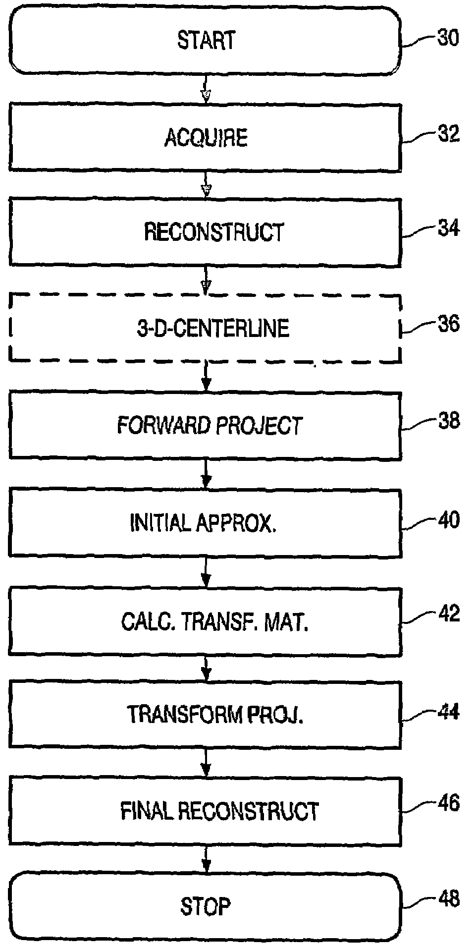
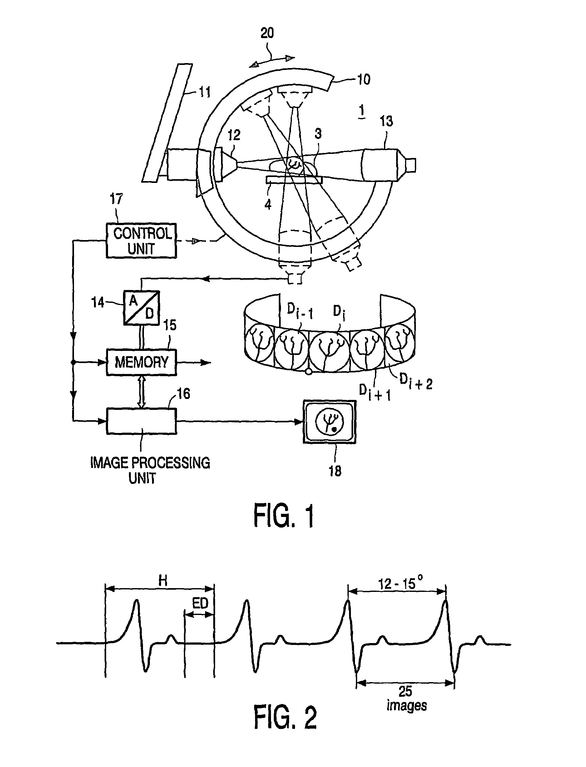
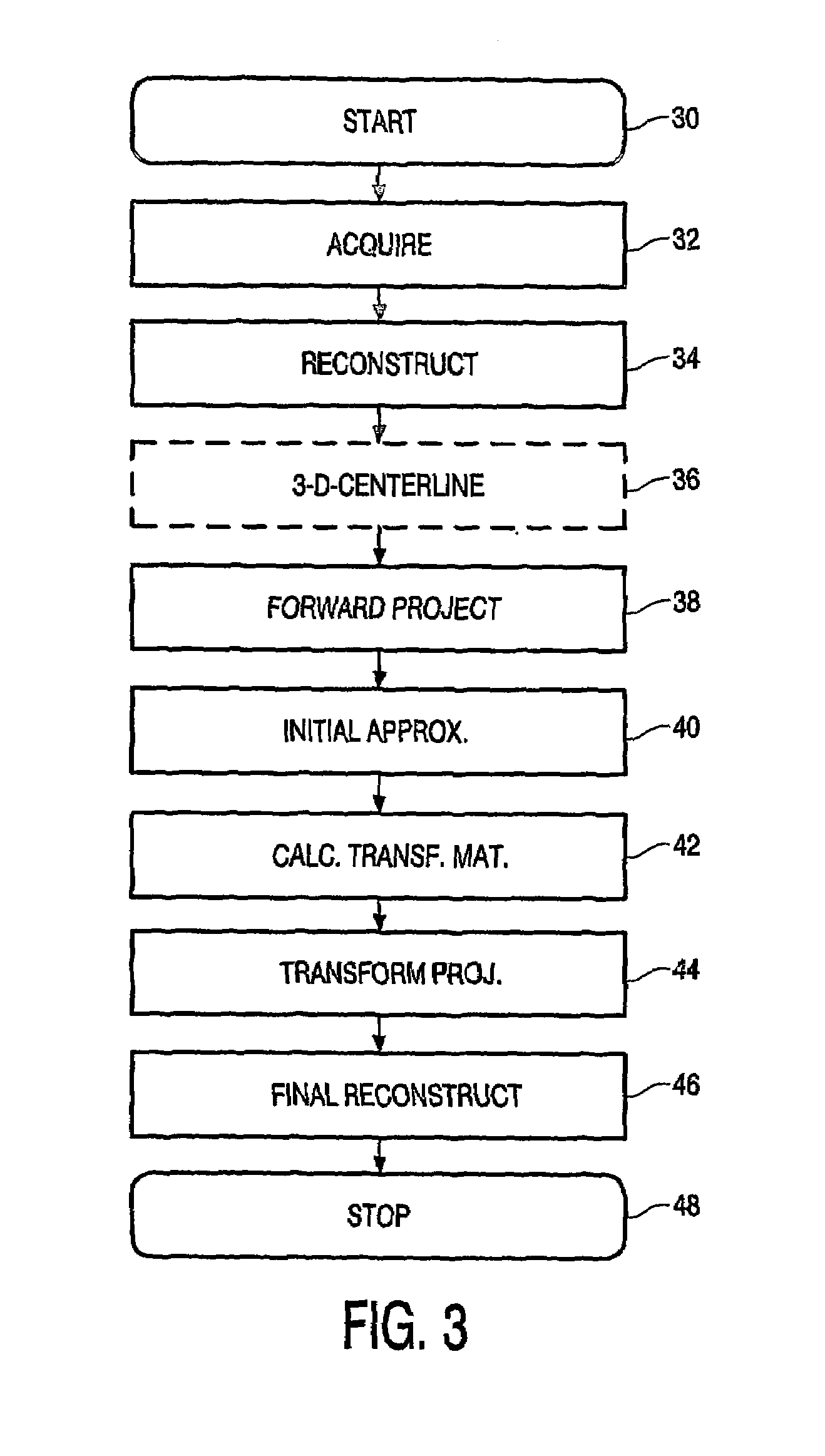
[0010]FIG. 1 illustrates an exemplary X-Ray imaging apparatus in which the invention may be applied. Basically, the apparatus allows to form two-dimensional X-Ray images of an object to be examined, in particular an object that moves (quasi-)periodically, such as a heart and its associated coronary vascular system, or alternatively, an object that moves rather unpredictably, such as an intestine or part thereof. From the combining of various such two-dimensional images, a three-dimensional volume of the object should be obtained. By itself, such three-dimensional reconstruction is state of the art. Similar technology may be used to produce a four-dimensional data set such as representing a selected part of the heart during a phase interval of its motion.

[0011]Now, the imaging apparatus 1 includes a C-arm 10 that is mounted on a partially shown stand 11. The C-arm can be rotated over an angle such as 180° around its center in the direction of double arrow a 20 through a motor drive n...


PUM

PropertyMeasurementUnit
angleaaaaaaaaaa
angleaaaaaaaaaa
angleaaaaaaaaaa

Abstract

An X-ray imaging method forms a set of a plurality of two-dimensional X-Ray projection images of a medical or veterinary object to be examined through a scanning rotation by an X-Ray source viz à viz the object. Such X-Ray images are acquired at respective predetermined time instants with respect to a functionality process produced by the object. From said set of X-Ray projection images by back-projection a three-dimensional volume image of the object is reconstructed. In particular, an appropriate motion correction is derived for the respective two-dimensional images, and subsequently as based on a motion vector field from the various corrected two-dimensional images the intended three-dimensional volume is reconstructed.

Description

BACKGROUND OF THE INVENTION[0001]The invention relates to an X-ray imaging method, comprising the forming of a set of a plurality of two-dimensional X-Ray projection images of a medical or veterinary object to be examined through a scanning rotation by an X-Ray source viz à viz said object, which X-Ray images are acquired at respective predetermined time instants with respect to a functionality process produced by said object, and from said set of X-Ray projection images reconstructing by back-projection a three-dimensional volume image of said object.[0002]Such methods have been in use for cardiac and other medical diagnostic operations. The prime problem for acquiring an appropriate 3D or 4D data set from the 2-dimensional images is the movement of items within the human or other body under consideration, which movement is sometimes substantially periodic, but not completely so. In other circumstances, the object in question, such as an intestine can be considered as moving in qui...

Claims


Application Information

Patent Timeline
Patent Type & Authority Patents(United States)
IPC IPC(8): A61B6/03A61B6/00G01N23/04G06T11/00
CPCA61B6/5264G01N23/046G06T11/005A61B6/00A61B6/508A61B6/541G06T2211/404G01N2223/612A61B6/503G01N2223/419G06T2211/412
Inventor RASCHE, VOLKERMOVASSAGHI, BABAKBOOSTEN, MARCEL
Owner KONINKLIJKE PHILIPS ELECTRONICS NV
Why Eureka
Try Eureka