System for combined transcutaneous blood gas monitoring and negative pressure wound treatment

a technology of blood gas monitoring and wound treatment, applied in the field of monitoring of blood gas, can solve the problems of increased risk of tissue injury, erythema, blisters, burns and skin tears, inappropriate treatment of patients, etc., and achieve the effect of reducing or eliminating contamination risk

Inactive Publication Date: 2009-04-28
KCI LICENSING INC
View PDF142 Cites 105 Cited by
  • Summary
  • Abstract
  • Description
  • Claims
  • Application Information

AI Technical Summary

Benefits of technology

[0014]The blood gas transducer unit is in fluid communication with the vacuum source through an interposed vacuum hose and in electrical communication with the blood gas data acquisition device through an interposed electrical cable. The vacuum source, which comprises a vacuum pump operated by a pump motor is placed in fluid communication with the blood gas transducer unit in order to induce a condition of hyperperfusion in the locality of the electrochemical blood has transducer. Under the control of the microcontroller, or equivalent means, the blood gas data acquisition device is then utilized to capture this measure to arrive at an estimate of arterial partial pressure of oxygen or carbon dioxide, accordingly. Because vacuum induced perfusion produces the requisite condition of hyperperfusion without local heating and, therefore, without acceleration of the local metabolic function, the present invention results in more accurate than previously available estimates of partial blood gas pressures and does so while eliminating a significant risk for injury to the patient.
[0016]Because the application of vacuum perfusion to the patient presents at least some risk for contamination of the vacuum source and blood gas data acquisition device, the preferred embodiment of the present invention further comprises a transducer interface module particularly adapted for the reduction or elimination of contamination risk. According to the invention, the transducer interface module comprises a male and female interface pair, wherein the male portion is adapted into the female portion and thereby establishes communication between the blood gas transducer unit and the vacuum source and blood gas data acquisition device.
[0017]In implementing the male plug, a hydrophobic membrane filter—known to those of ordinary skill in the art—is interposed in the vacuum hose, thereby eliminating the opportunity for contaminants to pass from the patient to the vacuum source or blood gas data acquisition device. While the preferred embodiment of the present invention comprises a throw-away male plug, vacuum hose, electrical cable and blood gas transducer unit, those of ordinary skill in the art will recognize that each of these components can be made reusable with implementation of proper, known sterilization techniques. In this latter case, the hydrophobic membrane filter is preferably replaceable.

Problems solved by technology

Unfortunately, such local heating carries with it an increased risk for tissue injury—erythema, blisters, burns and skin tears being among the documented complications.
In addition, some debate exists within the art as to whether the increased local metabolic rate concomitant the application of heat counteracts the intended perfusion effect.
If so, false readings may result, which may ultimately lead to inappropriate treatment of the patient.
As a result, operator time is generally wasted in the administration of a transcutaneous blood gas evaluation.
Additionally, transcutaneous blood gas monitors are either not available for emergency use or must be made available with an operated in a standby mode.
Such a standby mode requires additional hardware and generally shortens the electrode lifecycle.

Method used


Image

  • System for combined transcutaneous blood gas monitoring and negative pressure wound treatment
  • System for combined transcutaneous blood gas monitoring and negative pressure wound treatment
  • System for combined transcutaneous blood gas monitoring and negative pressure wound treatment

Examples


Embodiment Construction

[0023]Although those of ordinary skill in the art will readily recognize many alternative embodiments, especially in light of the illustrations provided herein, this detailed description is exemplary of the preferred embodiment of the present invention, the scope of which is limited only by the claims that may be drawn hereto.

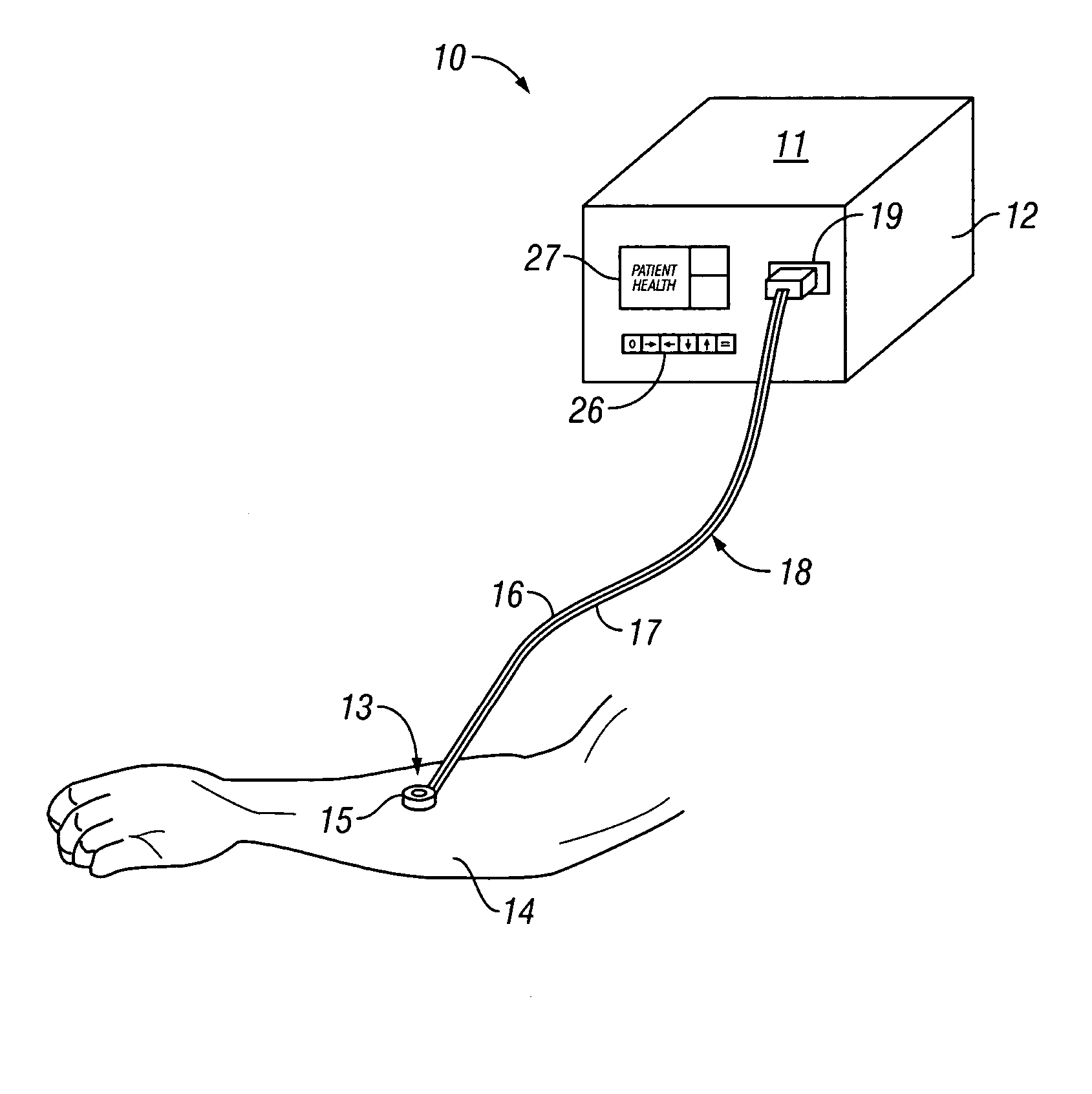
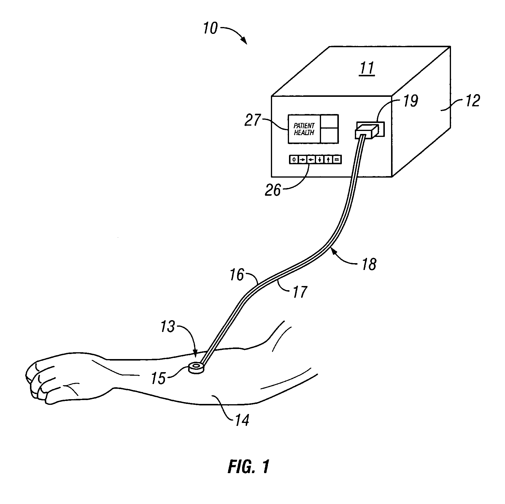
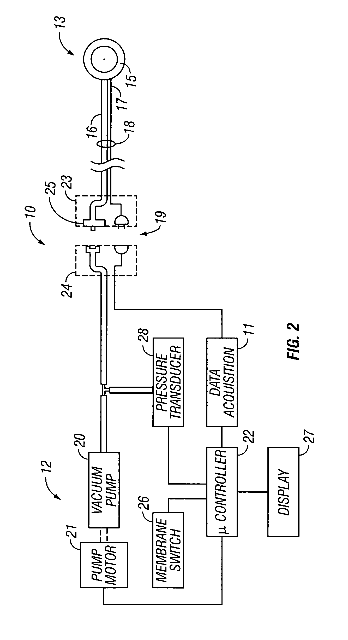
[0024]Referring now to FIG. 1, the preferred embodiment of the transcutaneous blood gas monitoring system 10 of the present invention is shown to generally comprise a blood gas data acquisition device 11, a vacuum source 12 and a blood gas transducer unit 13. As shown in FIG. 1, the blood gas transducer unit 13 is adapted for application to a patient's skin 14. In alternative embodiments, not shown, the blood gas transducer may be applied within a wound bed 30 or disposed within a screen 32 placed within the wound bed 30. As will be better understood further herein, the blood gas transducer unit 13 is also adapted for administration of a local vacuum at the are...


PUM


Abstract

A method and apparatus for the transcutaneous monitoring of blood gases generally comprises a blood gas data acquisition device, a vacuum source and a blood gas transducer unit. The blood gas transducer unit is adapted for application to a patient's skin and administration of a local vacuum at the area of patient application. It further comprises an electrochemical blood gas transducer, well known to those of ordinary skill in the art, which is disposed entirely within the local vacuum at the area of patient application. The vacuum source is placed in fluid communication with the blood gas transducer unit, through a hydrophobic membrane filter for safety purposes, in order to induce a condition of hyperperfusion in the locality of the electrochemical blood gas transducer. Under the control of a microcontroller, or equivalent means, the blood gas acquisition device is then utilized to capture a measure of skin surface oxygen or carbon dioxide pressure. The microcontroller can then utilize this measure to arrive at an estimate of arterial partial pressure of oxygen or carbon dioxide, accordingly. Because vacuum induced perfusion produces the requisite condition of hyperperfusion without local heating and, therefore, without acceleration of the local metabolic function, the present invention results in more accurate than previously available estimates of partial pressure blood gas pressures and does so while eliminating a significant risk for injury to the patient.

Description

RELATED APPLICATIONS[0001]This application is a continuation of U.S. patent application Ser. No. 10 / 085,321 filed Feb. 28, 2002 now U.S. Pat. No.6,856,821, which is a continuation-in-part, of U.S. patent application Ser. No. 09 / 579,755, entitled “Transcutaneous Blood Gas Monitoring with Vacuum Pefusion,” filed on May 26, 2000, now abandoned which claims priority from Provisional Application No. 60 / 136293, filed May 27, 1999.FIELD OF THE INVENTION[0002]The present invention relates to the monitoring of blood gases during vacuum assisted wound healing. More particularly, the invention relates to a method and system for the transcutaneous monitoring of blood gases wherein said monitoring is enhanced by application of a vacuum pressure in the region of skin under evaluation, and during which negative pressure therapy is being applied to an adjacent or proximal wound site.BACKGROUND OF THE INVENTION[0003]Transcutaneous blood gas monitoring is known in the relevant arts as a method by whi...

Claims


Application Information

Patent Timeline
Patent Type & Authority Patents(United States)
IPC IPC(8): A61B5/00A61M1/00
CPCA61B5/14542A61B5/1468A61B5/445A61B5/6834A61M1/0088A61M1/962A61M1/915
Inventor JOHNSON, ROYCE
Owner KCI LICENSING INC
Why Eureka
Try Eureka