Ultrasonic contrast agent detection and imaging by low frequency manipulation of high frequency scattering properties

a contrast agent and ultrasonic technology, applied in diagnostics, applications, reradiation, etc., can solve the problems of high transmission amplitude, low image resolution typical obtained with harmonic imaging, and the destruction of contrast agent bubbles, so as to increase the lateral and range resolution, increase the cnr, and increase the range resolution

Active Publication Date: 2010-01-05
ANGELSEN BJORN A J +1
View PDF16 Cites 15 Cited by
  • Summary
  • Abstract
  • Description
  • Claims
  • Application Information

AI Technical Summary

Benefits of technology

[0018]With non-moving, temporary stationary tissue, one can for example transmit two pulses with different phase of the low frequency components and the same phase of the high frequency components, and perform a linear combination of the back-scattered signals from the two pulses. The scattered high frequency components from the contrast bubble will be manipulated differently than from the tissue by the two low frequency pulses of different phases and / or amplitudes, and the bubble signal can be preserved while the tissue signal is heavily suppressed in the combination of the two echoes.
[0022]The present invention significantly increases the CNR relative to existing methods by using the total scattered high frequency signal, and in particular the strong linear components, from the contrast agent and not only nonlinear components of it.
[0023]Relative to nonlinear harmonic imaging methods, the present invention can use a more broadbanded transmit pulse and will hence achieve a higher range image resolution.
[0024]In addition, a higher transmit frequency can be used resulting in a significant increase in both lateral and range resolution relative to nonlinear imaging methods.
[0025]The performance of the present invention will not be sensitive to the amplitude of the imaging pulses compared to nonlinear imaging methods. Together with the indicated suppression of received tissue signal with resulting increase in CNR, this facilitates nondestructive detection and imaging of single contrast agent bubbles.

Problems solved by technology

The consequence of a relatively narrowbanded and low frequency drive pulse is the low image resolution typically obtained with harmonic imaging.
Also, the received nonlinear harmonic component from the contrast agent typically has low amplitude which reduces the CNR and may require so high transmitted amplitude that the contrast agent bubbles are destroyed.
This can cause a problem when the inflow rate of contrast agent to the tissue region is low.
Finally, a limitation in all methods based on nonlinear harmonic detection is that nonlinear components in the tissue signal is preserved in the process, also limiting the CTR.

Method used


Image

  • Ultrasonic contrast agent detection and imaging by low frequency manipulation of high frequency scattering properties
  • Ultrasonic contrast agent detection and imaging by low frequency manipulation of high frequency scattering properties
  • Ultrasonic contrast agent detection and imaging by low frequency manipulation of high frequency scattering properties

Examples


Embodiment Construction

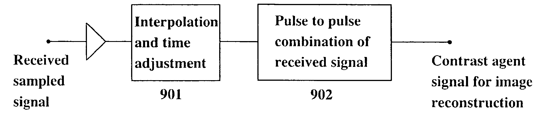
[0037]The invention will now be described in more detail with reference to the figures.

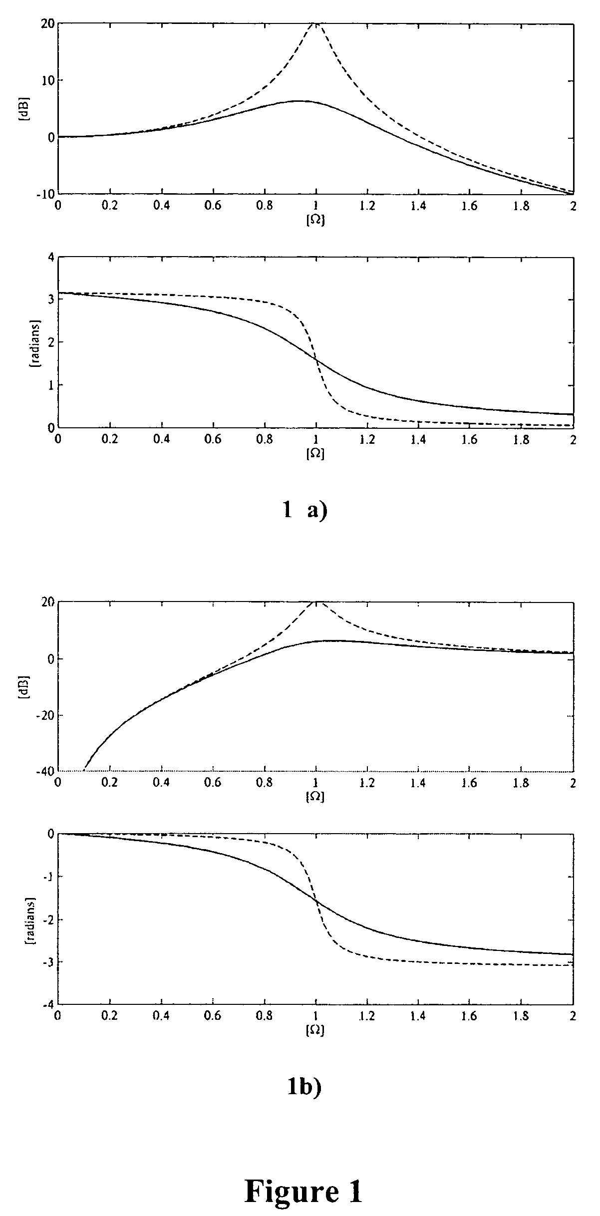
[0038]For small amplitude radius excursions, the mathematical equations governing contrast bubble oscillation can be linearized and we obtain the following transfer function from incident pressure on the bubble surface to radial bubble displacement

[0039]H1⁡(Ω)=1Ω2-1-i⁢⁢Ω⁢⁢d

where

[0040]d=bω0⁢m,ω02=sm,Ω=ωω0

[0041]Here, ω is the angular frequency and ω0 is the resonance frequency of the bubble while s is the stiffness of the gas and shell, m is the inertia of the surrounding liquid, and d is a damping factor of the resonant system.

[0042]The absolute value and phase angle of H1(Ω) is shown in the upper and lower panel in FIG. 1a, respectively. In the lower panel, we see that for drive frequencies well below resonance the displacement is π out of phase with the driving pressure. For frequencies well above resonance the bubble responds differently and the displacement and drive pressure are now in phase s...


PUM


Abstract

A method for improved detection and imaging of ultrasound contrast agents using dual-band transmitted pulses, is described. The method is based on transmitting a pulse consisting of two frequency bands, a low frequency band which purpose is to manipulate the high frequency scattering properties of the contrast agent, and a high frequency band from which the image reconstruction is based. In addition, a general form of pulse subtraction is used to significantly suppress the received tissue signal.

Description

CROSS REFERENCE TO RELATED APPLICATIONS[0001]This application is a continuation of U.S. patent application Ser. No. 10 / 851,820 filed May 21, 2004 now abandoned which claims priority from U.S. Provisional Patent Application Ser. No. 60 / 475,222 filed May 30, 2003.BACKGROUND OF THE INVENTION[0002]1. Field of the Invention[0003]This invention relates to methods and systems for ultrasonic detection and imaging of contrast agents located in soft tissue or tissue fluids.[0004]2. Description of the Related Art[0005]Ultrasound contrast agents are typically made as solutions of micro gas bubbles or nano lipid particles. The gas bubbles typically show strong and nonlinear scattering of the ultrasound, a phenomenon that is used to differentiate the contrast agent signal from the tissue signal. In the earliest applications (˜1985) the increased scattering from the contrast agent within the transmitted frequency band was used to enhance the scattering from blood. Later, second harmonic components...

Claims


Application Information

Patent Timeline
Patent Type & Authority Patents(United States)
IPC IPC(8): A61B8/00G01S7/52G01S15/89
CPCG01S7/52022G01S7/52026G01S15/8952G01S15/8963A61B8/481
Inventor ANGELSEN, BJORN A. J.HANSEN, RUNE
Owner ANGELSEN BJORN A J
Why Eureka
Try Eureka