Model-based grayscale registration of medical images

a technology of medical images and model data, applied in image enhancement, image analysis, instruments, etc., can solve the problems of reducing the likelihood of a total mastectomy, missing diagnosis, and prolonging life expectancy, so as to enhance the speed and reliability of cad detection

Active Publication Date: 2010-02-23
HOLOGIC INC
View PDF31 Cites 28 Cited by
  • Summary
  • Abstract
  • Description
  • Claims
  • Application Information

AI Technical Summary

Benefits of technology

[0015]According to a preferred embodiment, numerical image processing algorithms are performed on two or more medical images to provide grayscale registration thereof, the numerical image processing algorithms being based at least in part on a model of medical image acquisition. The grayscale registered temporal images may then be displayed and / or further processed for comparison thereof. Methods and systems grayscale registration of two or more medical images such as mammograms for facilitating visual comparison thereof and enhancing the speed and reliability of CAD detection of medical abnormalities are disclosed. It should be appreciated that the present invention can be implemented in numerous ways, including as a process, an apparatus, a system, a device, a method, or a computer readable medium such as a computer readable storage medium or a computer network wherein program instructions are sent over optical or electronic communication lines. Several inventive embodiments of the present invention are described below.
[0017]The various acquisition models described herein take into account many of the most relevant steps in the image acquisition that influence pixel value differences between images. The parameter-based model provides a transform function or lookup table TA,B(gA) to transform or register pixel values in one image to those on another image. The methods are particularly suited for temporal comparisons of medical images such as mammograms. In addition, the transformation facilitates comparison of analog with digital images, i.e., combinations of unprocessed digital and digitized film-based mammograms by a radiologist. The method is based on a model of acquisition, which makes it relatively unsusceptible to differences between the images that are not due to acquisition, while other differences do not greatly affect the quality of transformed images negatively.
[0018]The method enables a radiologist to make quick, effective and efficient temporal comparisons of medical images and thus requiring fewer manual and subjective adjustments to be made by the radiologist for optimal display of the mammograms. In addition or alternative to image display, the transformation method may also be used for computer aided detection methods that make use of temporal other comparative information.

Problems solved by technology

While not a replacement for the experienced radiologist, CAD systems are designed to increase efficiency and reduce error, as a typical radiologist may be required to examine hundreds of medical images per day, which can lead to the possibility of a missed diagnosis due to human error.
While at present there is no means for preventing breast cancer, early detection of the disease prolongs life expectancy and decreases the likelihood of the need for a total mastectomy.
However, in conventional radiology the review of temporal image pairs may be seriously hampered by differences in acquisition.
These differences may cause subsequent mammograms to appear dramatically different and thus reduce the effectiveness of temporal comparisons.
Thus, for example, when visually comparing a current-year mammogram to a prior-year mammogram on a softcopy display, a radiologist can have difficulty detecting subtle anatomical differences between the mammograms, because their different acquisition parameters can make them “look” very different in terms of grayscale levels.
These differences may not be easily normalized as they may induce non-linear gray scale changes.
Although radiologists may adjust the relative contrast, brightness and / or position of the threshold of the S-curve (the HD-curve, named after Hurter and Driffeld) of one or more images, such a process can be inaccurate, tedious, annoying, and time-consuming and may reduce performance and induce human errors.
In other words, these acquisition differences cannot be easily or quickly corrected by the radiologists during display.
In addition to negatively affecting visual comparison thereof, differences in grayscale settings / parameters between two temporally distinct mammograms of the same breast can be a disadvantage for temporal comparisons thereof in a CAD system, such as that described in U.S. Pat. No. 6,075,879 supra.

Method used


Image

  • Model-based grayscale registration of medical images
  • Model-based grayscale registration of medical images
  • Model-based grayscale registration of medical images

Examples


Embodiment Construction

[0040]The following description is presented to enable any person skilled in the art to make and use the invention. Descriptions of specific embodiments and applications are provided only as examples and various modifications will be readily apparent to those skilled in the art. The general principles defined herein may be applied to other embodiments and applications without departing from the spirit and scope of the invention. Thus, the present invention is to be accorded the widest scope encompassing numerous alternatives, modifications and equivalents consistent with the principles and features disclosed herein. For purpose of clarity, details relating to technical material that is known in the technical fields related to the invention have not been described in detail so as not to unnecessarily obscure the present invention.

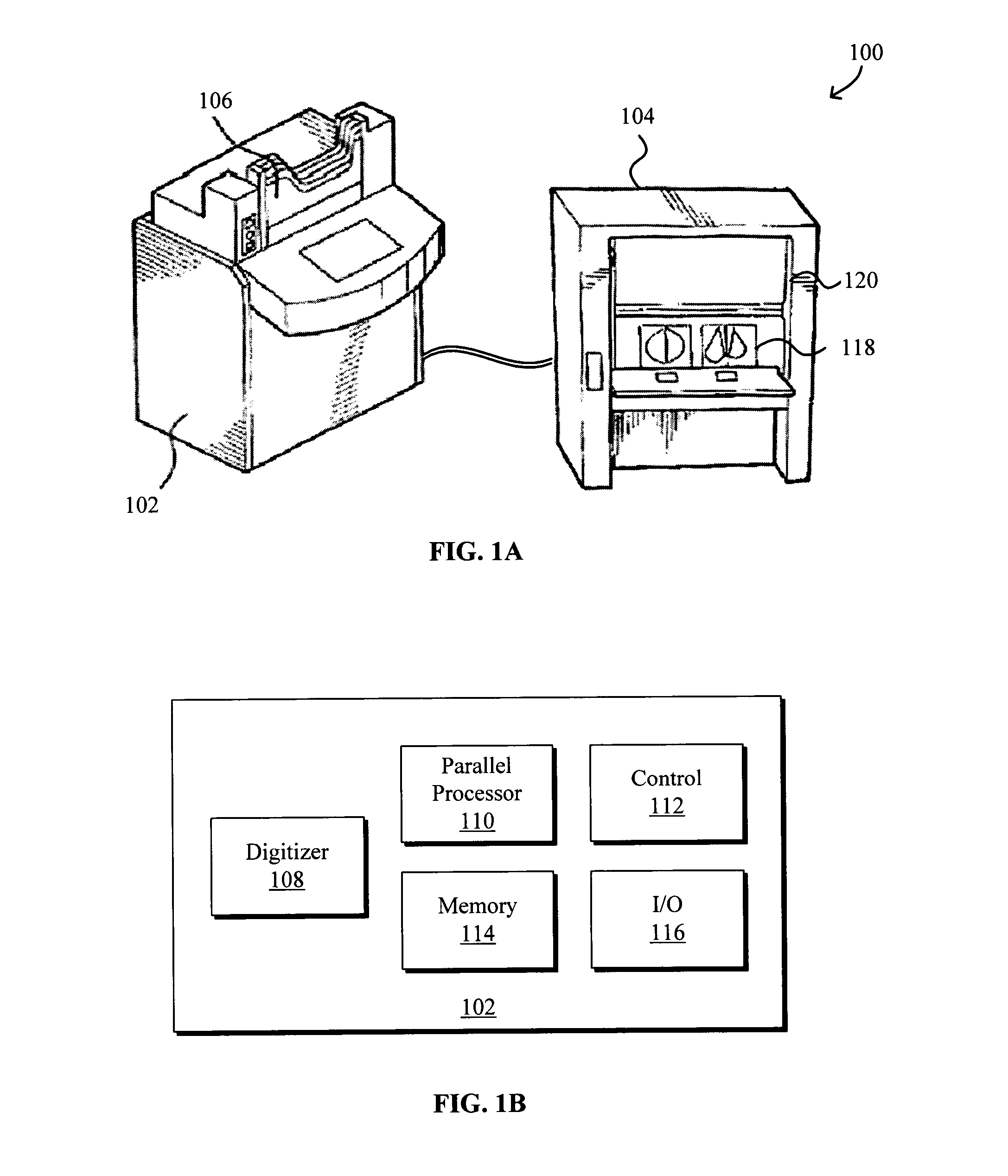
[0041]FIG. 1A shows an outside view of a computer aided diagnostic (CAD) system 100, such as an IMAGE CHECKER M1000 from R2 Technology, Inc., for assisting ...


PUM


Abstract

Numerical image processing based on a model of medical image acquisition of two or more medical images to provide grayscale registration is described. The grayscale registered temporal images may then be displayed for visual comparison and / or further processed by a computer-aided diagnosis system for detection of medical abnormalities therein. A parametric method includes spatially registering two images and performing gray scale registration of the images. A parametric transform model, e.g., analog to analog, digital to digital, analog to digital, or digital to analog model, is selected based on the image acquisition method(s) of the images, i.e., digital or analog / film. Gray scale registration involves generating a joint pixel value histogram from the two images, statistically fitting parameters of the transform model to the joint histogram, generating a lookup table, and using the lookup table to transform and register pixel values of one image to the pixel values of the other image.

Description

CROSS REFERENCE TO RELATED APPLICATION[0001]The subject matter of this application is generally related to the subject matter of commonly assigned, U.S. patent application Ser. No. 10 / 622,978, entitled “Facilitating Computer-Aided Diagnosis, Comparison, And / Or Display Of Medical Images” and filed concurrently on Jul. 18, 2003, which was published on Oct. 28, 2004 as US2004 / 0213447A1, the entirety of which is incorporated herein by reference.BACKGROUND OF THE INVENTION[0002]1. Field of the Invention[0003]This patent specification relates generally to display and / or computer aided diagnosis (CAD) of medical images. More specifically, methods and systems grayscale registration of two or more medical images such as mammograms for facilitating visual comparison thereof and enhancing the speed and reliability of CAD detection of medical abnormalities are disclosed.[0004]2. Description of Related Art[0005]Systems for computer-aided diagnosis (“CAD”) assist radiologists in the detection and...

Claims


Application Information

Patent Timeline
Patent Type & Authority Patents(United States)
IPC IPC(8): G06K9/00G06K9/32G06T7/00
CPCG06T7/0034G06T2207/30068G06T7/35
Inventor SNOEREN, PETER R.KARSSEMEIJER, NICO
Owner HOLOGIC INC
Why Eureka
Try Eureka