Devices, systems and methods for detecting increase fluid levels in tissue

a technology of fluid level and tissue, applied in the field of devices, systems and methods of detecting increased fluid level in tissue, can solve the problems of increasing the risk of extravasation, affecting the safety of patients, and causing serious injury to patients

Active Publication Date: 2010-03-09
BAYER HEALTHCARE LLC
View PDF31 Cites 32 Cited by
  • Summary
  • Abstract
  • Description
  • Claims
  • Application Information

AI Technical Summary

Problems solved by technology

Extravasation can be caused, for example, by rupture or dissection of fragile vasculature, valve disease, inappropriate needle placement, or patient movement resulting in the infusing needle being pulled from the intended vessel or causing the needle to be pushed through the wall of the vessel.
High injection pressures and / or rates of some modem procedures can increase the risk of extravasation.
Extravasation can cause serious injury to patients.
In that regard, certain injection fluids such as contrast media or chemotherapy drugs can be toxic to tissue.
Such plethysmographs can be cumbersome to operate and / or insensitive to small changes in volume.
Use of electrodes in impedance plethysmographs can, however, result in instabilities.
For example, maintaining suitable electrical (ohmic or galvanic) contact between the electrodes of impedance plethysmographs and the skin of the patient is often very difficult.
Because light is heavily absorbed in tissue, however, the sensitivity of photo-plethysmographs is generally limited to the top ¼ inch of tissue.
Moreover, the injection medium may flow into interstitial spaces remote from the photoplethysmograph sensors and go undetected.
Although measurement of temperature changes and emissivity using microwave energy can result in instantaneous detection, temperature differences are often too small for practical measurement.

Method used


Image

  • Devices, systems and methods for detecting increase fluid levels in tissue
  • Devices, systems and methods for detecting increase fluid levels in tissue
  • Devices, systems and methods for detecting increase fluid levels in tissue

Examples


Embodiment Construction

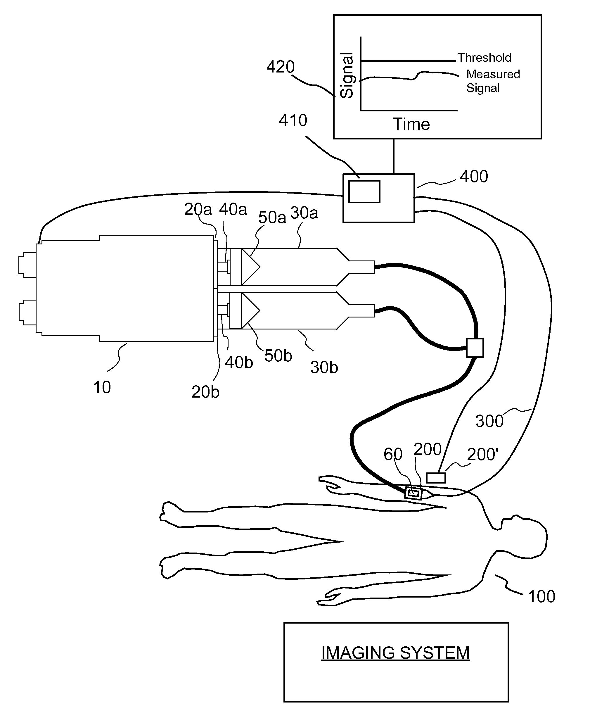
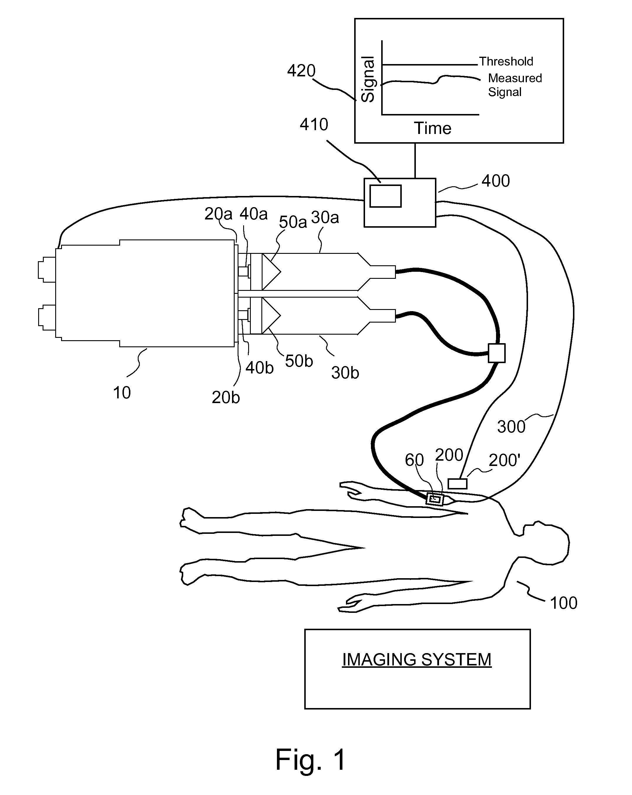
[0033]In several embodiments, the present invention provides a closed-loop technique for determining the stability of an injection site by administering a relatively nontoxic, harmless or benign first fluid (for example, saline) to a patient while monitoring the injection site with an extravasation sensor. This administration step occurs prior to the injection of a second fluid, for example, a therapeutic or diagnostic bolus of a drug. If the first fluid (for example, saline) is determined to extravasate into the tissue, the first fluid is less toxic than the second fluid to be injected and is likely to cause complications as a result of the extravasation.

[0034]As illustrate in FIG. 1, one embodiment of an injection system of the present invention includes an injector 10 such as the STELLANT® injector available from Medrad, Inc. of Indianola, Penn. Injector 10 includes two syringe interfaces 20a and 20b to which two syringes 30a and 30b are removably attachable. Two drive members or...


PUM


Abstract

A system for injection of a fluid into a body includes: a source of a first fluid, a source of a second fluid; the first fluid being less toxic than the second fluid; at least one pressurizing system in operative connection with the source of the first fluid and with the source of the second fluid; at least one controller in operative connection with the pressurizing system; and at least a first sensor in communicative connection with the controller. The first sensor is adapted to transmit a signal of a detected change in fluid level in tissue indicative of extravasation to the controller. The controller is adapted to cause injection of the first fluid into the body to determine if extravasation of the first fluid occurs using the sensor in an administration phase before injection of the second fluid.

Description

BACKGROUND OF THE INVENTION[0001]The present invention relates generally to the devices, systems and methods of detecting increased fluid levels in tissue, and particularly to devices, systems and method of detecting increased fluid levels associated with extravasation in tissues and controlling powered injectors in response to such detection.[0002]The following information is provided to assist the reader to understand the invention disclosed below and the environment in which it will typically be used. The terms used herein are not intended to be limited to any particular narrow interpretation unless clearly stated otherwise in this document. References set forth herein may facilitate understanding of the present invention or the background of the present invention. The disclosure of all references cited herein are incorporated by reference.[0003]Elevated fluid levels in tissue can arise as a result of introduction of a fluid into the body, for example, during an injection procedu...

Claims


Application Information

Patent Timeline
Patent Type & Authority Patents(United States)
IPC IPC(8): A61M1/00A61B6/00
CPCA61M5/14546A61M5/16827A61M5/14566A61M2005/1588
Inventor KALAFUT, JOHNEZEKIEL, GUY
Owner BAYER HEALTHCARE LLC
Why Eureka
Try Eureka