Mechanically registered videoscopic myringotomy/tympanostomy tube placement system

a technology of tympanostomy and tympanostomy tube, which is applied in the field of mechanical registration of videoscopic myringotomy/tympanostomy tube placement system, systems, devices, etc., can solve the problems of increasing the risk and cost of tympanostomy tube placement, the risk and cost of out-patient surgical procedures performed under general anesthesia, and the significant portion of the risk and cost of tympanosto

Inactive Publication Date: 2010-04-27
TUSKER MEDICAL
View PDF31 Cites 77 Cited by
  • Summary
  • Abstract
  • Description
  • Claims
  • Application Information

AI Technical Summary

Benefits of technology

[0009]The present invention provides improved devices, systems, methods, and kits for treating the tissue structures of the ear. The invention often makes use of a guide structure that can mechanically register a treatment probe with a target region of the tympanic membrane or eardrum. Mechanical registration may be provided by a structure which is fittingly received in an external auditory canal of the ear. The guide structure will often include a conformable body (typically comprising a compressible foam, or the like) so as to allow the guide structure to accommodate a range of differing auditory canal anatomy. The guide structure may further include an articulating mechanism for selectively orienting the treatment probe toward the target region of the tympanic membrane. The articulating mechanism will often selectively orient a probe lumen, with the treatment probe having a shaft fittingly sliding in the probe lumen so that engagement between a positioning surface of the guide structure and a tissue surface of the patient's ear maintains registration of the treatment probe. The guide structure may also support a videoscopic image capture device, illumination transmitting optical fibers, an aiming beam transmitter, and the like. Advantageously, such structures facilitate performing treatment procedures such as myringotomy, tympanostomy tube placement, and the like, under local (rather than general) anesthesia, often in a doctor's office (rather than an out-patient surgical facility).
[0012]The guide structure may be registered by articulating a treatment lumen of the guide structure relative to a positioning surface of the guide structure. For example, the guide structure may comprise a shaft eccentrically carrying the treatment lumen. The shaft may rotate within the auditory canal about an axis with the treatment probe precessing about the axis so as to orient the probe toward the target region. The positioning surface of the guide structure may be disposed over the shaft with a bearing therebetween to facilitate rotation without injury to the tissue surface engaged by the positioning surface. The shaft may flex during rotation so as to accommodate a bend of the auditory canal. The probe and / or other components of the treatment system within the guide structure may likewise flex during rotation.
[0020]The system will preferably include aiming means for identifying an orientation of the treatment probe relative to the tympanic membrane. In many embodiments, a light beam (such as from a laser or light-emitting diode) may be directed onto the tympanic membrane at a location aligned with the treatment probe. Alternative embodiments may make use of a reticule superimposed on the image of the tympanic membrane as shown in a monitor to the system operator, a template superimposed on the image to indicate the target region, or the like. Such aiming structures can verify registration of the treatment probe with the target region before, during, and / or after piercing the tympanic membrane, tube deployment, and the like.
[0022]A limit surface of the treatment probe may engage a limit surface of the guide structure or the tympanic membrane so as to inhibit axial movement of the shaft after the probe pierces the tympanic membrane. This can help avoid inadvertent injury to the middle and / or inner ear structures or undesired positioning of the tube distally of the tympanic membrane within the middle ear. In the exemplary embodiment, engagement of the limit surfaces inhibits movement when the distal end of the probe advances from the guide structure by a distance in a range from about 1.0 mm to about 40 mm.

Problems solved by technology

In recent years, more complex and expensive systems have been proposed for diagnosis or treatment of the tissues of the ear, including systems using laser energy for forming a myringotomy, video systems for imaging of the ear canal, and the like.
In particular, there are both risks and costs associated with out-patient surgical procedures performed under general anesthesia.
For example, a significant portion of the risk and cost of tympanostomy tube placement is associated with the administration of general anesthesia, i.e., the need for an operating room, the presence of an anesthesiologist, and related recovery room time.

Method used


Image

  • Mechanically registered videoscopic myringotomy/tympanostomy tube placement system
  • Mechanically registered videoscopic myringotomy/tympanostomy tube placement system
  • Mechanically registered videoscopic myringotomy/tympanostomy tube placement system

Examples


Embodiment Construction

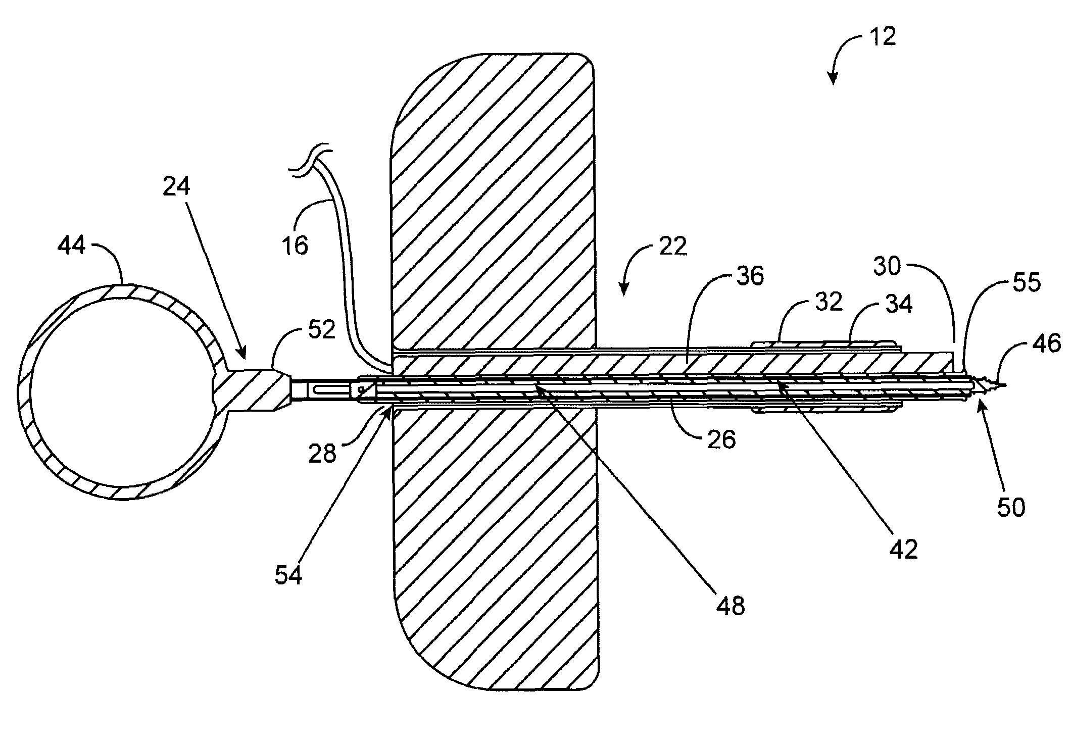
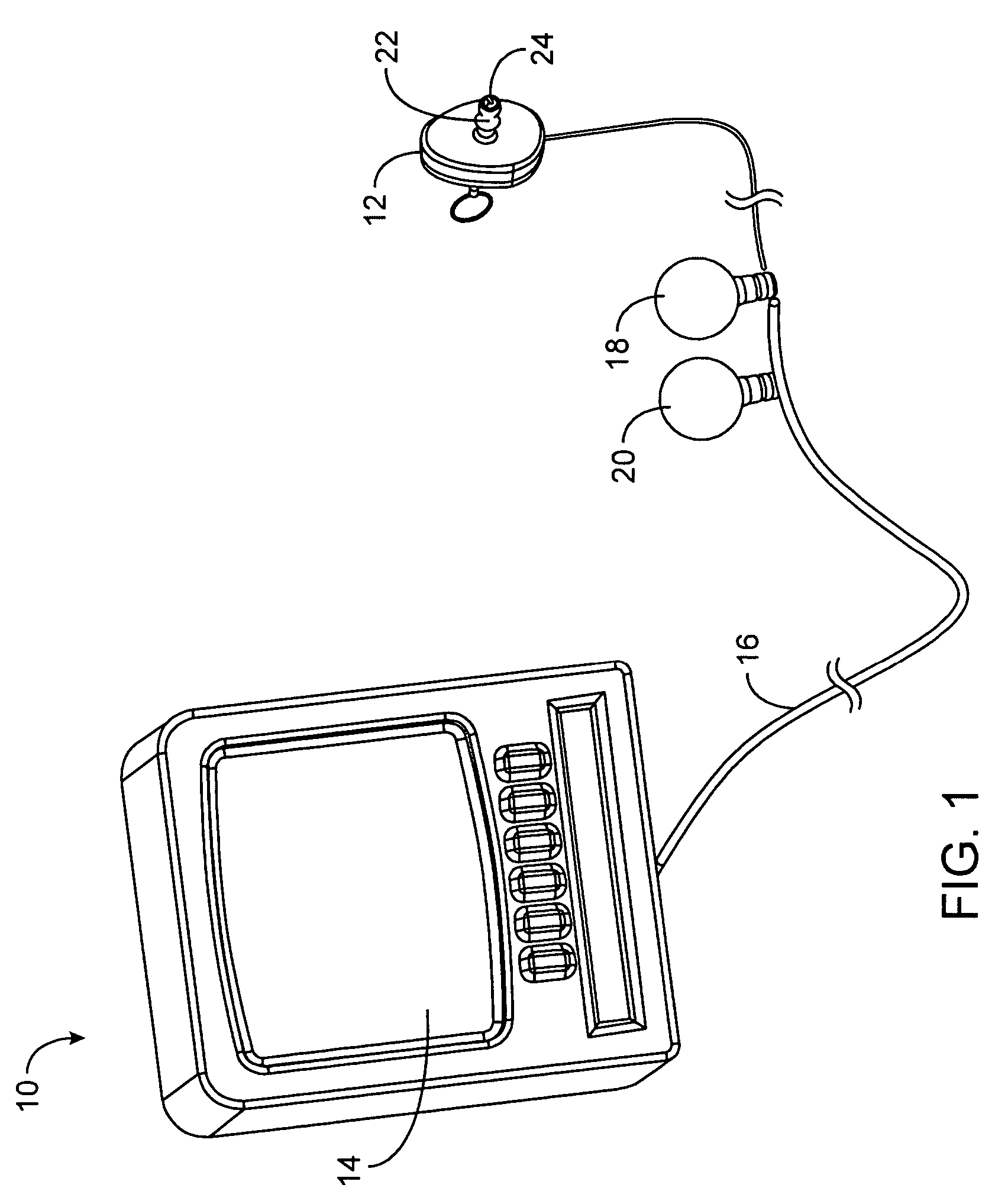
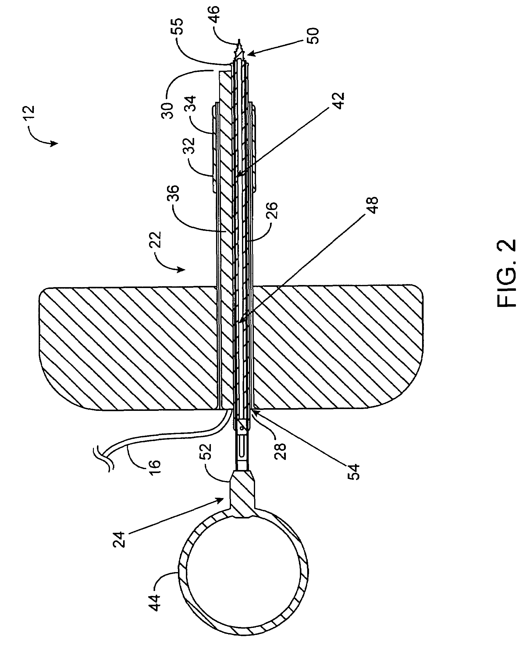
[0039]The present invention generally provides improved devices, systems, methods, and kits for treatment of tissue structures of the ear. Myringotomy, tympanostomy tube placement, and other procedures may be performed using a guide structure to register one or more treatment probes relative to the target tissue structure. The guide structure will often be articulatable, allowing selective registration of the treatment probe with a target region of, for example, the tympanic membrane or eardrum. The guide structure may be supported by, for example, a conformable body, such as a compressible foam insertable into the external auditory canal. Engagement between the conformable body and the auditory canal can maintain a position of the guide structure so that the guide structure, in turn, maintains an orientation of the treatment probe. Such a lightweight guide structure may be mountable to the patient, allowing stabilized videoscopic imaging from an image capture device supported by th...


PUM


Abstract

Devices, systems, methods, and kits for treating the tissue structures of the ear make use of a guide structure that can mechanically register a treatment probe with a target region of a target tissue, the guide structure being fittingly received in an auditory canal and often comprising a conformable body such as a compressible foam, or the like. The guide structure may include an articulating mechanism for selectively orienting the treatment probe toward the target region of, for example, a tympanic membrane. The guide structure may also support a videoscopic image capture device, illumination transmitting optical fibers, an aiming beam transmitter, and the like. Such structures facilitate myringotomy, tympanostomy tube placement, and the like, under local anesthesia in a doctor's office.

Description

CROSS-REFERENCE TO RELATED APPLICATION[0001]This patent application is a divisional application which claims priority from U.S. patent application Ser. No. 09 / 843,541 filed Apr. 26, 2001, now U.S. Pat. No. 6,770,080, the full disclosure of which is incorporated herein by reference.BACKGROUND OF THE INVENTION[0002]The present invention is generally related to medical devices and apparatus. In particular, the invention provides systems, methods, devices, and kits for treating a patient's ear. In one embodiment, the invention provides a system and method for myringotomy with or without tympanostomy tube placement.[0003]Otitis media is among the most common diagnosis made by pediatricians. A majority of children may have at least one episode of otitis media (“earache”) prior to their third birthday. Otitis media is often caused by an inability of the eustachian tube to drain fluid from the middle ear. Otitis media is often treated with antibiotics.[0004]A significant number of children ...

Claims


Application Information

Patent Timeline
Patent Type & Authority Patents(United States)
IPC IPC(8): A61F11/00
CPCA61F11/002A61B90/30A61B90/37A61F11/202A61F11/20
Inventor KAPLAN, AARON V.TARTAGLIA, JOSEPHVAUGHAN, ROBERTJONES, CHRISTOPHER
Owner TUSKER MEDICAL
Why Eureka
Try Eureka