
Actuation mechanism for flexible endoscopic device

a flexible endoscope and actuator technology, applied in the field of medical devices, can solve the problems of inability to allow an endoscopist to both feed and operate, delay in procedure, incomplete tissue removal, etc., and achieve good end effector actuation and enhance the ability of the flexible shaft.

Inactive Publication Date: 2010-05-04
ETHICON ENDO SURGERY INC
View PDF32 Cites 49 Cited by
  • Summary
  • Abstract
  • Description
  • Claims
  • Application Information

AI Technical Summary

Benefits of technology

Enables the endoscopist to both feed and operate endoscopic accessories independently, reducing miscommunication and maintaining effective end effector function even in tortuous paths, thereby improving procedure efficiency and tissue sampling.

Problems solved by technology

For example, navigating a tortuous bending colon or introducing a gastroscope into an esophagus can be a difficult and time-consuming part of a procedure.
These existing designs do not allow an endoscopist to both feed and operate (e.g. slide, open, close, actuate, etc.) the accessory being used with the endoscopist's single free hand.
Although this procedure using an assistant is used, there may be delays or miscommunication between endoscopist and assistant as to when or where to operate an accessory that results in procedure delays, misdiagnosis, or incomplete tissue removal.
Another issue that occasionally arises when using an endoscopic accessory is that winding or otherwise positioning a long, flexible accessory instrument in a tortuous path can result in a reduction in ability to open or close the end effector at the distal end of the device.
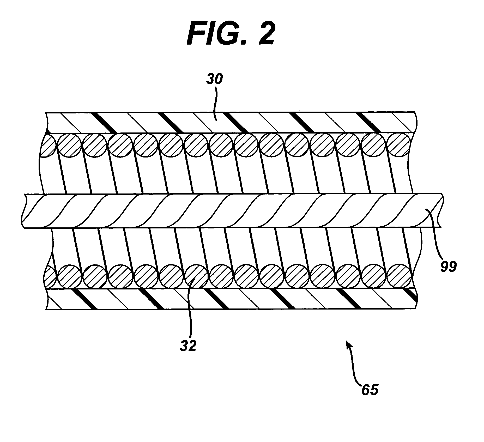
This loss in the ability to open or close the device results from the free floating pull cable (typically inside a long flexible device) being placed in tension as the shaft is wound, causing the end effector to partially close independently of actuation of the handle.
Such limitations in the end effector motion may reduce the ability to perform a procedure, or reduce the force with which jaws close, affecting an operator's ability to sample tissue adequately.

Method used


Image

  • Actuation mechanism for flexible endoscopic device
  • Actuation mechanism for flexible endoscopic device
  • Actuation mechanism for flexible endoscopic device

Examples


Embodiment Construction

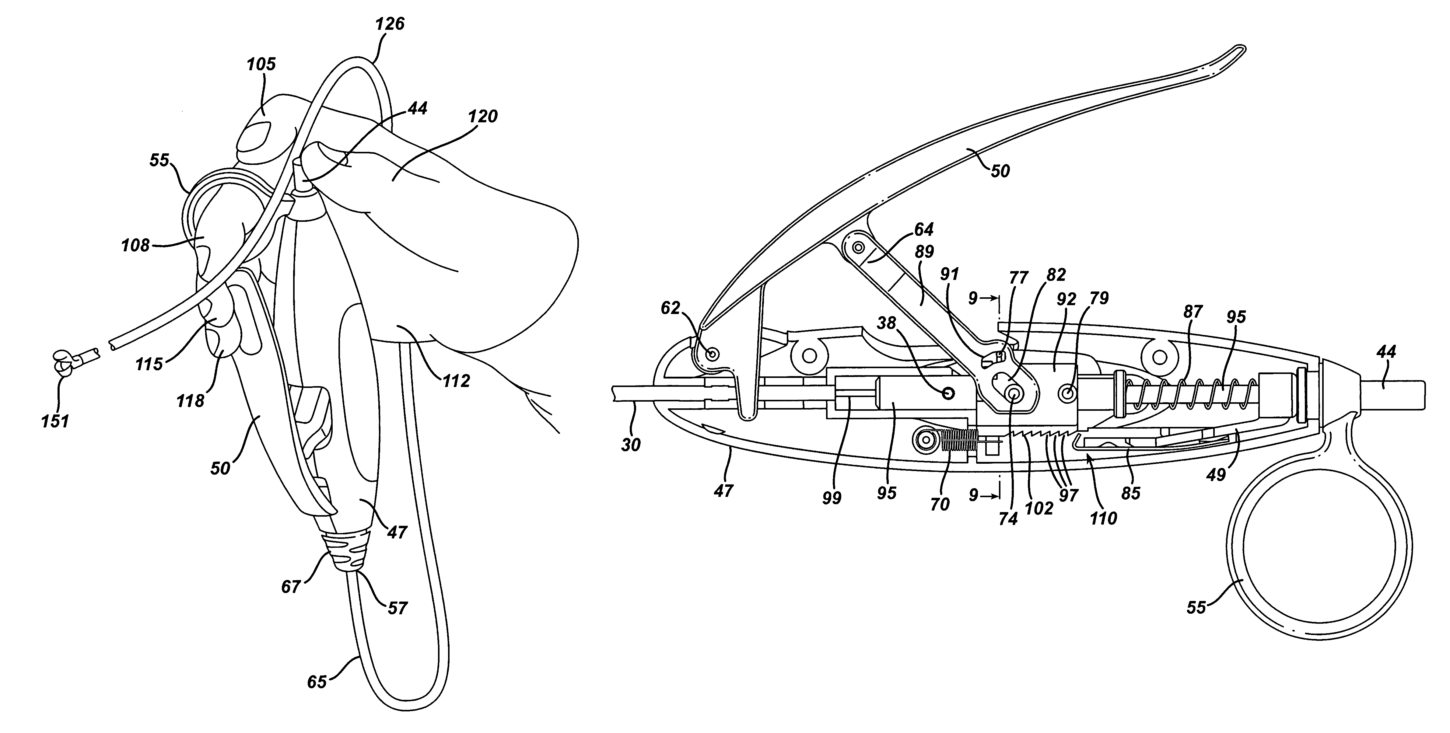
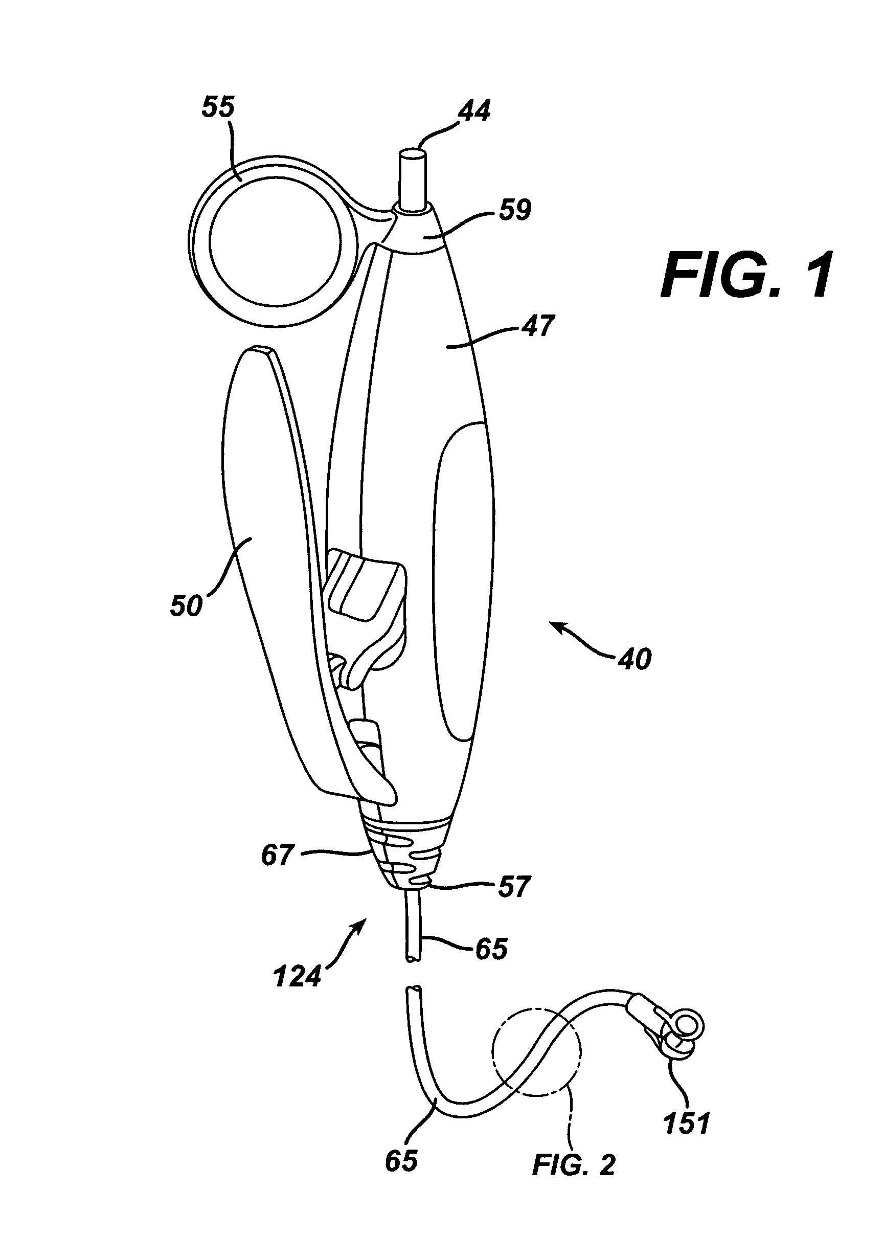
[0029]FIG. 1 shows a novel medical device handle 40 according to the present invention associated with the proximal end of an endoscopic accessory instrument 124. The accessory 124 illustrated in FIG. 1 includes a biopsy jaw pair 151 (also referred to as biopsy jaws 151) at the distal end of the accessory 124. For illustrative purposes, the description that follows uses biopsy jaws 151 as an example of a suitable end effector on endoscopic accessory 124, but it is apparent to those skilled in the art that handle 40 may be used with other accessory instruments having other end effectors or other devices positioned at the distal end of accessory 124 for providing diagnostic and / or therapeutic function(s), such as, but not limited to, biopsy forceps such as biopsy jaws 151, grasping forceps, surgical scissors, extractors, washing pipes and nozzles, needle injectors, non energized snares, and electosurgical snares.

[0030]FIGS. 15A-15I illustrate various end effectors. FIG. 15A illustrate...


PUM


Abstract

An endoscopic accessory medical device is provided. The device can include a handle, a flexible shaft, and an end effector. The handle can include an actuator for operating the end effector through a wire or cable pulling member that extends through the flexible shaft. The handle and actuator can be operable with a single hand, such that the operation of the end effector can be accomplished with the same hand that is used to hold the handle and advance the end effector through an endoscope. The handle can include an actuation mechanism that is decoupled from operation of the end effector when the actuator is in a first open position, which becomes operatively coupled to the end effector when the actuator is moved to a second position, such as by squeezing the actuator, and which operates the end effector when the actuator is moved further to a third position.

Description

FIELD OF THE INVENTION[0001]The present invention relates, in general, to medical devices, and more particularly to handles on endoscopic and surgical accessories.BACKGROUND OF THE INVENTION[0002]Endoscopists typically perform diagnosis and therapy using a flexible endoscope such as a gastroscope, colonoscope, enteroscope, cystoscope, or other types of endoscopes. The endoscope provides the endoscopist with the ability to visualize the inside of a lumen, and is often designed with an integral working channel through which small accessory devices may be passed to perform therapy at various tissue sites within a body.[0003]Guiding the flexible endoscope to the desired location within a body lumen requires a high level of skill. For example, navigating a tortuous bending colon or introducing a gastroscope into an esophagus can be a difficult and time-consuming part of a procedure. Therefore, much of the endoscopist's skills are related to using and handling the endoscope. Unlike certai...

Claims


Application Information

Patent Timeline
Patent Type & Authority Patents(United States)
IPC IPC(8): A61B17/28A61B10/06A61B1/00A61B10/00A61B17/00A61B17/32A61B17/34
CPCA61B10/06A61B17/2909A61B17/3201A61B17/3478A61B17/32056A61B2017/00477A61B2017/00292A61B2017/00367A61B1/00
Inventor NOBIS, RUDOLPHHESS, CHRISTOPHER J.STOKES, MICHAEL J.
Owner ETHICON ENDO SURGERY INC
Learn More