
Systems and methods for automated diagnosis and grading of tissue images

a tissue image and automatic diagnosis technology, applied in image enhancement, image data processing, instruments, etc., can solve the problems of subjective processes, affecting the normal arrangement of gland units, and patients with grade 5 tissue typically have a lower chance of survival

Inactive Publication Date: 2010-07-20
CHAMPALIMAUD FOUND +2
View PDF50 Cites 69 Cited by
  • Summary
  • Abstract
  • Description
  • Claims
  • Application Information

AI Technical Summary

Benefits of technology

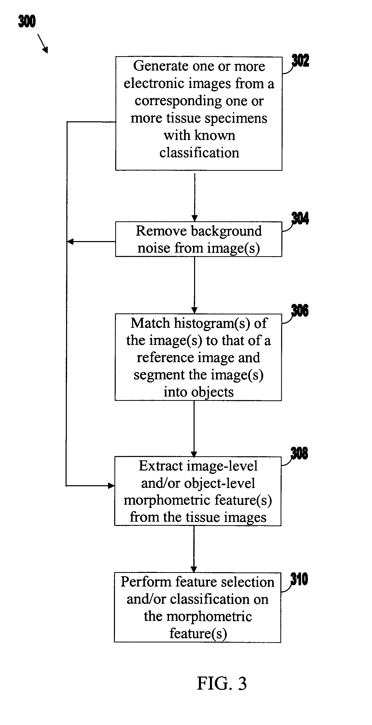
[0023]In another aspect of the present invention, systems and methods are provided that use a 2-stage procedure for tissue image analysis. In a first stage, a first set of morphometric data from a tissue image with unknown classification is evaluated by a model that predicts whether the corresponding tissue is cancerous or non-cancerous. In a second stage, if the model classifies the tissue as cancerous tissue, a second set of morphometric data from the tissue image is evaluated by a model that assigns a cancer grade to the tissue (e.g., a Gleason grade for prostate tissue). The second set of morphometric feature(s) may include at least one morphometric feature not included in the first set of morphometric feature(s). For example, in the first stage, one or more image-level morphometric features from the tissue image (e.g., fractal dimension, fractal code, wavelet, and / or color channel histogram feature(s)) may be evaluated by the model that predicts whether the tissue is cancerous or non-cancerous. In the second stage, one or more object-level features, alone or in combination with image-level features, may be evaluated by the model that assigns a cancer grade to the tissue. In an embodiment, the second set of morphometric features may be extracted from the tissue image only if the tissue is classified by the model as being cancerous. In this way, the resources of physicians, other individuals and / or automated processing equipment (e.g., equipment for extracting morphometric data from tissue images) may be conserved. In another embodiment, the first and second sets of morphometric features may be extracted from the tissue image at the same time. This two-level procedure may be used to, for example, identify in a whole tissue section (or other suitable size tissue section), portions of the section that are cancerous, and then to further analyze those sections.

Problems solved by technology

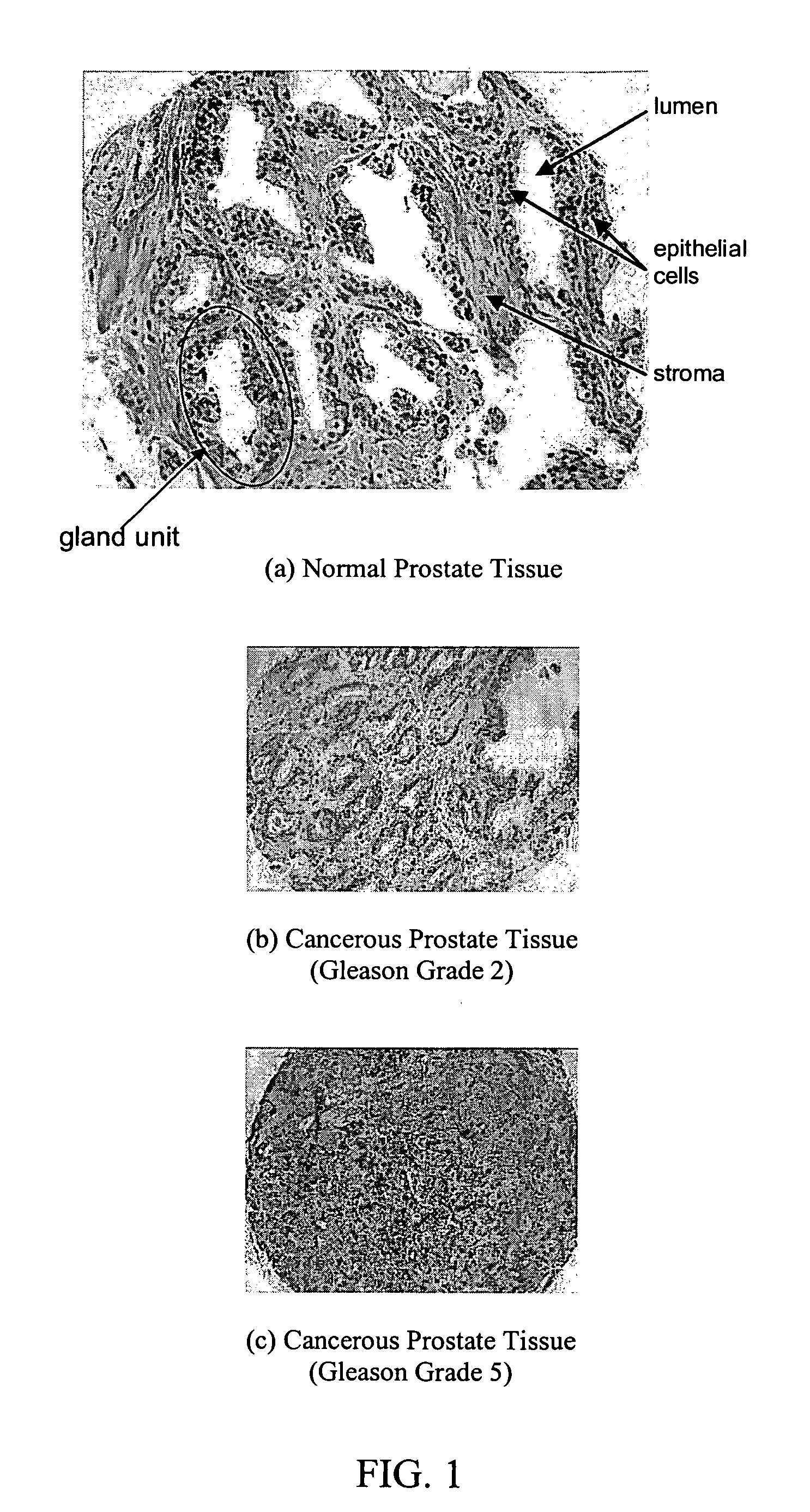
When cancer occurs, epithelial cells replicate in an uncontrolled way, thereby disrupting the normal arrangement of the gland units.
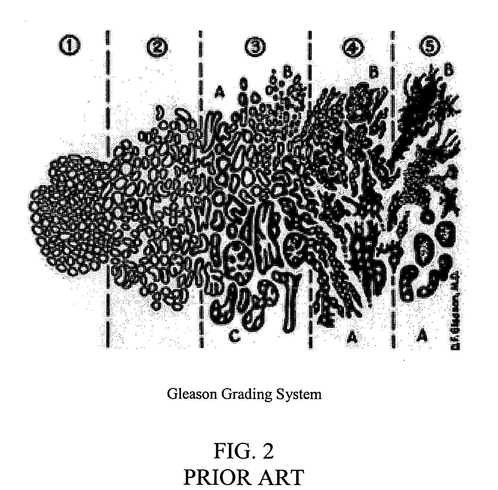
On the other hand, grade 5 corresponds to poorly differentiated tissue, and thus patients with grade 5 tissue typically have a lower chance of survival.
Although the above-described cancer diagnosis and Gleason grading by a pathologist are widely considered to be reliable, these are subjective processes.
Thus, different pathologists viewing the same tissue samples may come up with conflicting interpretations.
However, it should be noted that both of these systems involve image segmentation prior to extracting image features, which can introduce segmentation error.
This could potentially have introduced positive bias into the reported results.

Method used


Image

  • Systems and methods for automated diagnosis and grading of tissue images
  • Systems and methods for automated diagnosis and grading of tissue images
  • Systems and methods for automated diagnosis and grading of tissue images

Examples


Embodiment Construction

[0044]Embodiments of the invention relate to systems and methods for automated diagnosis and grading of tissue images. The diagnosis and / or grading may be based on any suitable image-level morphometric data extracted from the tissue images including fractal dimension data, fractal code data, wavelet data, and / or color channel histogram data. As used herein, “data” of a particular type (e.g., fractal dimension or wavelet) may include one or more features of that type. The diagnosis and / or grade may be used by physicians or other individuals to, for example, select an appropriate course of treatment for a patient. The following description focuses primarily on the application of the present invention to cancer diagnosis and Gleason grading of images of prostate tissue. However, the teachings provided herein are also applicable to, for example, the diagnosis, prognosis, and / or grading of other medical conditions in tissue images such as other types of disease (e.g., epithelial and mixe...


PUM


Abstract

Systems and methods are provided for automated diagnosis and grading of tissue images based on morphometric data extracted from the images by a computer. The morphometric data may include image-level morphometric data such as fractal dimension data, fractal code data, wavelet data, and / or color channel histogram data. The morphometric data may also include object-level morphometric data such as color, structural, and / or textural properties of segmented image objects (e.g., stroma, nuclei, red blood cells, etc.).

Description

CROSS-REFERENCE TO RELATED APPLICATIONS[0001]This claims the benefit of U.S. Provisional Patent Application Ser. Nos. 60 / 600,764, filed Aug. 11, 2004, and 60 / 645,158, filed Jan. 18, 2005, which are hereby incorporated by reference herein in their entireties.FIELD OF THE INVENTION[0002]Embodiments of the invention relate to systems and methods for automated diagnosis and grading of tissue images. For example, in one embodiment, the invention provides systems and methods for extracting morphometric data from an image of prostate tissue and evaluating the data to determine whether the tissue is cancerous or non-cancerous. In another embodiment, the invention evaluates the morphometric data to determine a grade of cancer aggressiveness (e.g., a Gleason grade) for the prostate tissue.BACKGROUND OF THE INVENTION[0003]Prostate cancer is the most prevalent form of cancer and the second most common cause of death among men in the United States. One of the most reliable methods for prostate c...

Claims


Application Information

Patent Timeline
Patent Type & Authority Patents(United States)
IPC IPC(8): G06F19/00G06K9/00G06K9/20G06K9/36
CPCG06K9/0014G06T7/0012G06T2207/30024G06V20/695
Inventor SAIDI, OLIVIERTABESH, ALITEVEROVSKIY, MIKHAIL
Owner CHAMPALIMAUD FOUND
Learn More