
Method for double row fixation of tendon to bone

a technology of tendon and bone fixation, applied in the field of arthroscopic surgery, can solve the problems of knot tying during surgery, tedious and time-consuming, knot deformation or collapse,

Active Publication Date: 2011-09-06
ARTHREX
View PDF8 Cites 71 Cited by
  • Summary
  • Abstract
  • Description
  • Claims
  • Application Information

AI Technical Summary

Benefits of technology

The present invention provides a system and method for repairing soft tissue to bone using a suture anchor and a knotless fixation device. The method involves creating a first medial row and a second lateral row with the fixation devices, and then securing a structure over the soft tissue using the anchors. The technical effect of this invention is to provide a more efficient and effective way to repair tendon to bone fixation.

Problems solved by technology

Surgical methods utilizing suture anchors alone are disadvantageous for reattachment of large areas of detached tissue because they often do not allow good tissue to bone contact.
Knot tying during surgery, particularly arthroscopic surgery, is tedious and time-consuming.
There is also a tendency for the knots to deform or collapse as the surgeon manually forces the knots down into the proper position.
Also, the suture knots often are exposed to abrasion or cutting by sharp or rough areas along the walls of the bone canal into which anchors are typically inserted to provide fixation of tendon to bone.

Method used


Image

  • Method for double row fixation of tendon to bone
  • Method for double row fixation of tendon to bone
  • Method for double row fixation of tendon to bone

Examples


Embodiment Construction

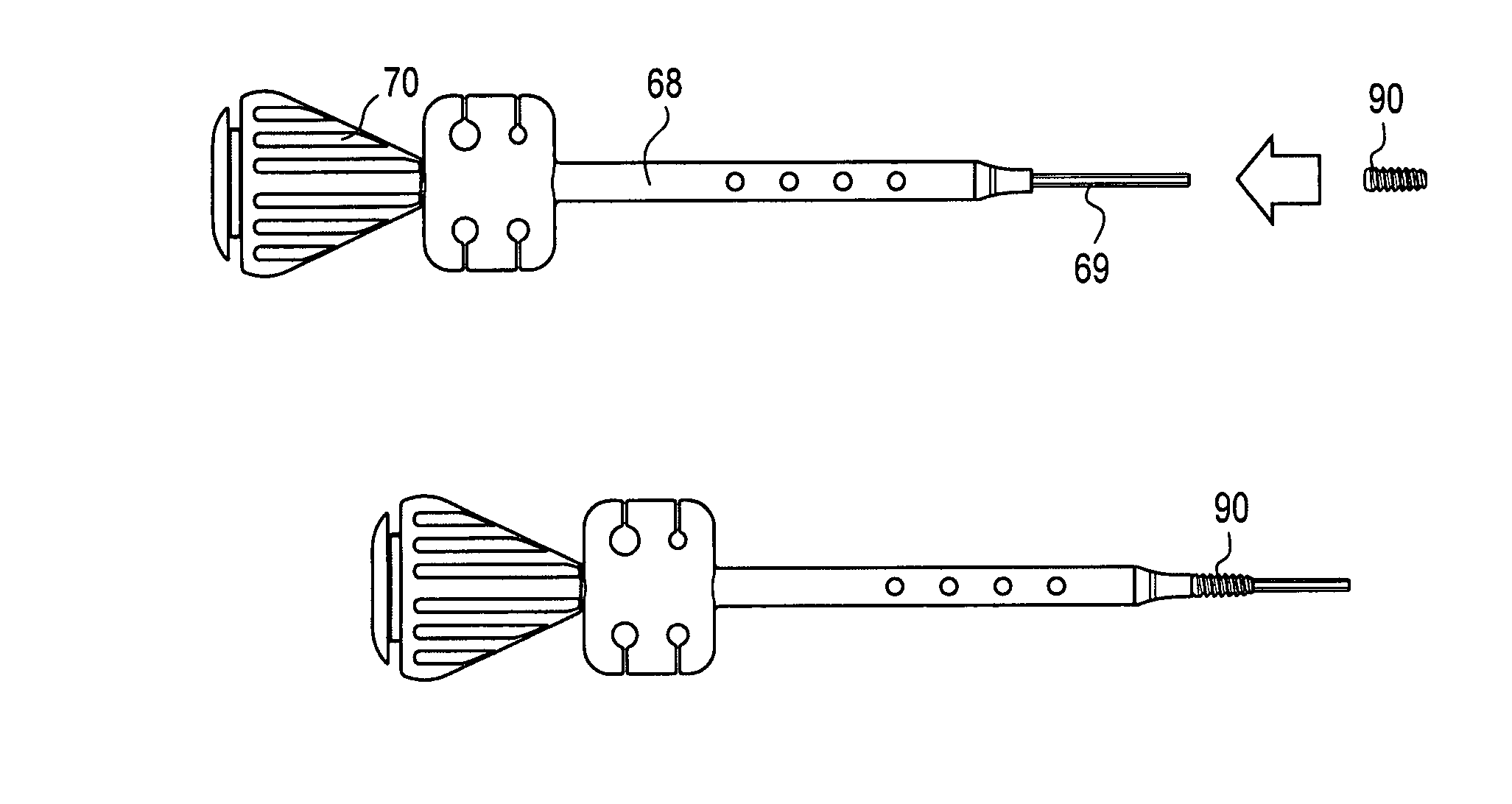
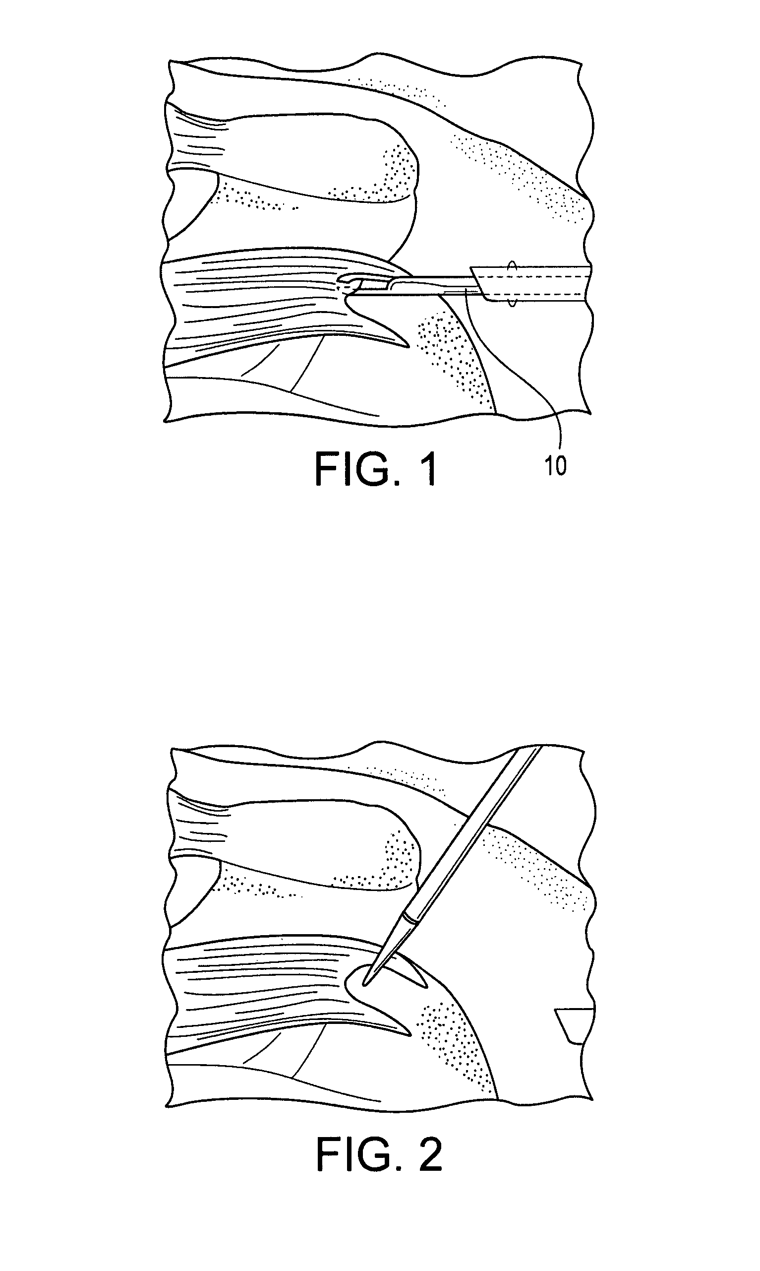
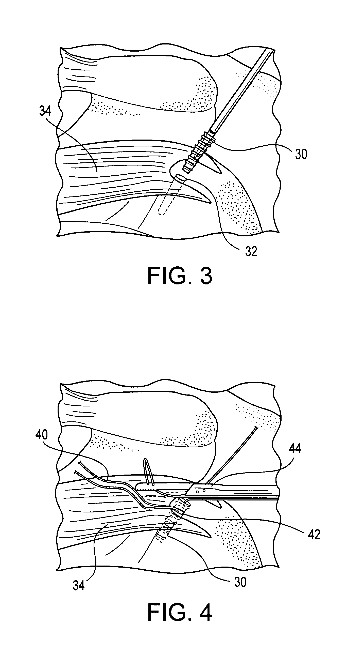
[0024]Referring now to the drawings, where like elements are designated by like reference numerals, FIGS. 1 through 15 illustrate systems and methods of attaching a tendon to bone according to the present invention. For exemplary purposes only, the invention will be described below with reference to an arthroscopic rotator cuff repair. However, the invention is not limited to this exemplary embodiment and has applicability to any reattachment of soft tissue to bone.

[0025]The methods of the present invention enhance footprint compression and allow for accelerated tendon healing to bone that is achieved with minimal knot tying. The repair consists of a tied medial row constructed with at least one suture anchor combined with knotless lateral fixation using at least one knotless fixation device. Preferably, the repair consists of a tied medial row constructed with two suture anchors (such as two Arthrex 5.5 mm Bio-Corkscrew® FT anchors, for example) combined with knotless lateral fixat...


PUM


Abstract

A system and method for soft tissue to bone repair employing at least one suture anchor combined with at least one knotless fixation device. The method for soft tissue to bone fixation includes: (i) providing a first medial row constructed with a first plurality of fixation devices, at least one of the first plurality of fixation devices is an anchor; and (ii) providing a second lateral row constructed with a second plurality of fixation devices, at least one of the second plurality of fixation devices is a knotless fixation device, and suture or tape or allograft / biological component extending over the soft tissue and secured in place by the anchors in the first and second medial rows.

Description

CROSS-REFERENCE TO RELATED APPLICATIONS[0001]This application claims the benefit of U.S. Provisional Application No. 60 / 763,915, filed Feb. 1, 2006, the entire disclosure of which is incorporated by reference herein.FIELD OF THE INVENTION[0002]The present invention relates to methods of arthroscopic surgery and, more specifically, to an improved method of attaching tissue to bone, such as rotator cuff repair.BACKGROUND OF THE INVENTION[0003]When soft tissue tears away from bone, reattachment becomes necessary. Various devices, including sutures, screws, staples, wedges, anchors and plugs have been used in the prior art to secure soft tissue to bone. Surgical methods utilizing suture anchors alone are disadvantageous for reattachment of large areas of detached tissue because they often do not allow good tissue to bone contact.[0004]Reattachment of soft tissue to bone typically requires the surgeon to pass suture material through selected tissue, form a plurality of surgical knots ext...

Claims


Application Information

Patent Timeline
Patent Type & Authority Patents(United States)
IPC IPC(8): A61B17/04A61F2/08
CPCA61B17/0401A61F2/0811A61B2017/0409A61B2017/044A61B2017/0414
Inventor ELATTRACHE, NEAL S.TIBONE, JAMES E.
Owner ARTHREX
Learn More