Methods for bone regeneration using endothelial progenitor cell preparations

a technology of endothelial progenitor cells and bone regeneration, which is applied in the field of bone regeneration using endothelial progenitor cell preparations, can solve the problems of limited body sites, delayed union or non-union at the fracture site, and too large defects for the body's natural healing respons

Active Publication Date: 2012-10-30
HEPACORE LTD
View PDF34 Cites 0 Cited by
  • Summary
  • Abstract
  • Description
  • Claims
  • Application Information

AI Technical Summary

Benefits of technology

[0032]In a preferred embodiment the step of administering the cell preparation comprises applying said cell preparation locally to the site of bone defect. Local administration assures that the administered EPCs remain in the desired area, thereby allowing a direct and rapid effect of the cells on the injured tissue.

Problems solved by technology

However, in approximately 10% of all fractures, the defect is too large for the body's natural healing response, and delayed unions or non-unions develop at the fracture site (Bancroft and Mikos, 2001; Rozen et al., 2007).
However, this process has several drawbacks including: limited body sites from which bone may be harvested without loss of function; autografts are less effective in irregularly shaped defects; and the procedure may be associated with complications such as infection, pain and nerve injury.
However, un-processed grafts carry a risk of disease transmission and immune rejection, while demineralized bone matrix is poor in bone growth inducing factors.
Xenografts are also a poor option due to the danger of disease transmission or rejection.
Metal implants may be permanently placed in bone to fill a defect however corrosion, infection and poor implant-tissue interface create many problems.
While offering excellent biocompatibility, they are often brittle and degrade too slowly thereby inhibiting natural bone re-growth.
As outlined above, the most advanced treatments are limited in effectiveness and are often associated with complications.

Method used


Image

  • Methods for bone regeneration using endothelial progenitor cell preparations
  • Methods for bone regeneration using endothelial progenitor cell preparations
  • Methods for bone regeneration using endothelial progenitor cell preparations

Examples


example 1

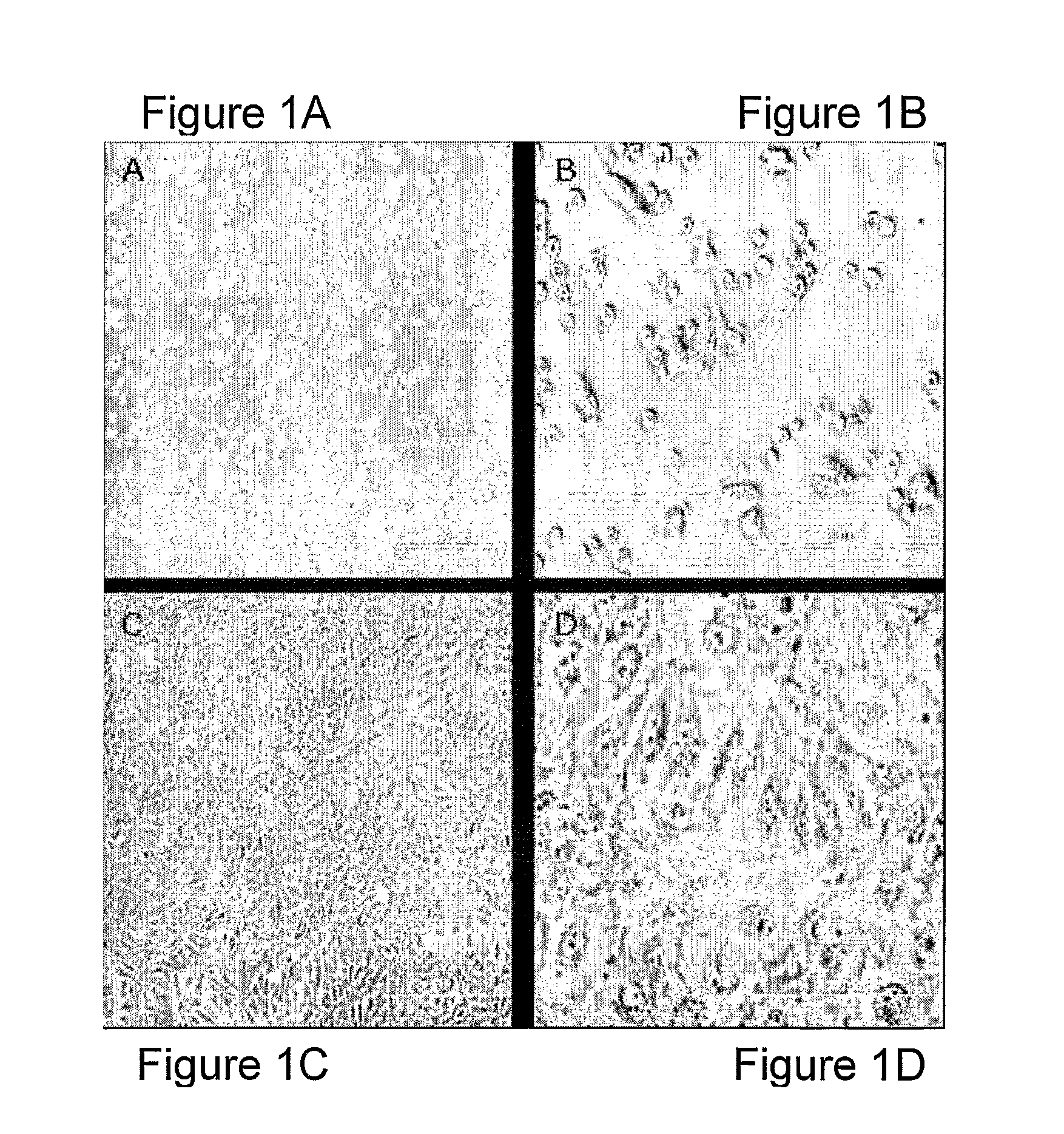
Ex-Vivo Expansion and Characterization of EPC

Isolation and Culture of EPC from Bone Marrow Derived and Peripheral Blood Derived Mononuclear Cells (MNC)

[0105]EPC were prepared from sheep mononuclear cells (MNC). Mononuclear cells (MNC) were separated from the bone-marrow of a removed tibia segment (3.2 cm) or from samples of 10 to 40 ml of peripheral venous blood taken from the jugular vein, using Uni-SepMAXI® U-16 Ficoll™ (Novamed, Jerusalem, Israel) by density gradient centrifugation at 400×g for 30 min at room temperature (RT). The MNC fraction was collected, washed twice, by centrifugation at 200×g for 15 min, with 45 ml Dulbecco's phosphate-buffered saline (DPBS, without calcium and magnesium; Biological Industries Ltd., Beit Haemek, Israel) containing 3% fetal calf serum (FCS; Biological Industries Ltd., Beit Haemek, Israel), and plated on fibronectin-coated six-well plates (Sigma Chemical Co., St. Louis, Mo.). Cells were cultured in endothelial basal medium-2 (EBM-2; Clonetics...

example 2

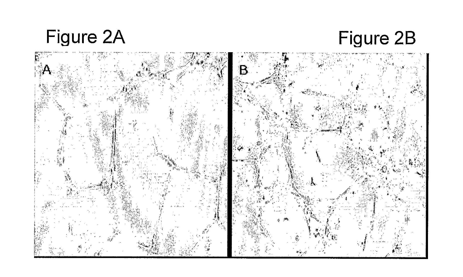
A Critical Gap Model in Sheep Tibiae

[0111]A critical bone gap model was generated by removing a 3.2 cm bone segment from sheep tibia (FIG. 5). Anesthesia was performed in 13 sheep (aged about 2 years, 40-70 Kilograms) by 0.4 xylasin and 600 mg ketamin, induced by 200 mg propapol and maintained by an intubation of 2% isofloran and oxygen ventilation. One gm cephazoline was given during surgery and then amoxilin 6 ml twice a day for the next 10 days. A longitudinal incision was performed along the skin of the anterior aspect of the right lower leg. The periosteum was similarly cut and elevated. Following the adjustment of a metal plate by 8 screws to the posterior aspect of the tibia, a segment of 3.2 cm was removed from the mid-diaphysis. This segment was preserved on ice for future isolation and culture of autologous EPC (as described above). Periosteum, fascia and skin were replaced and closed by sutures and pins and sprayed with antibiotics. A plaster cast was placed over the tibi...

example 3

Implantation of EPC or Paste into a Critical Bone Gap

[0112]Preparation of Amorphous Calcium Phosphate Paste

[0113]1.3 gr of Hydroxyapatite (ProChon Biotech) was dissolved in 10 ml of phosphate buffered saline (PBS). Following filtration, 2.5 gr of Hyaluronic acid (MW 3×10⁶ in 1% concentration, ProChon Biotech) were added.

[0114]Amorphous Calcium Phosphate Paste Containing FGF

[0115]1.3 gr of Hydroxyapatite (ProChon Biotech) was dissolved in 10 ml of phosphate buffered saline (PBS). 25 μgr / ml FGF2(3,5Q)-N111G (an FGF2 variant disclosed in WO 03 / 094835 assigned in part to the assignee of the present invention) was precipitated on the Hydroxyapatite for 1 hr at 37° C. Following filtration, 2.5 gr of Hyaluronic acid (MW 3×10⁶ in 1% concentration, ProChon Biotech) were added.

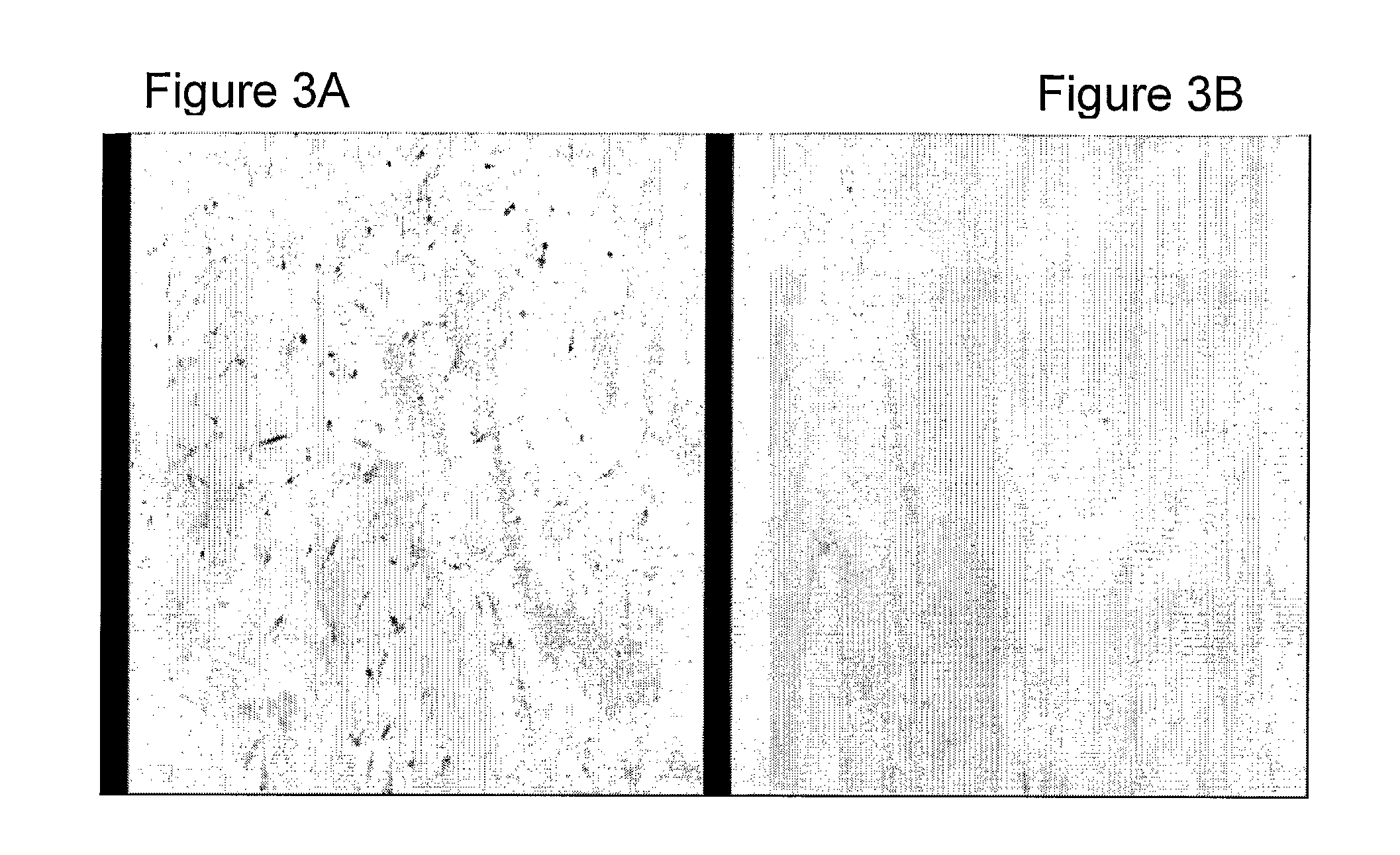
[0116]Two weeks after the removal of the bone segment, the sheep underwent a second operation using the same procedures. A nicely enveloped healing tissue was observed filling the whole segmental gap. A longitudinal wed...


PUM

PropertyMeasurementUnit
concentrationsaaaaaaaaaa
areaaaaaaaaaaa
conductiveaaaaaaaaaa

Abstract

The present invention relates in general to orthopedics and to a method for promoting repair of large bone defects, in particular non-union or delayed union fractures. Specifically the invention concerns the use of endothelial progenitor cell preparations for bone repair.

Description

[0001]This application is a 371 filing of International Patent Application PCT / IL2007 / 000485 filed Apr. 17, 2007, which claims the benefit of application No. 60 / 792,341 filed Apr. 17, 2006.FIELD OF THE INVENTION[0002]The present invention relates to a method for treating bone defects, in particular for promoting repair of large bone defects, including non-union and delayed union fractures. Specifically the invention concerns compositions comprising endothelial progenitor cells and methods of use thereof for bone repair.BACKGROUND[0003]Bone is a unique type of tissue composed of an inorganic mineral phase and cellular and extracellular matrix phases. Bone is a vital organ that undergoes modeling and remodeling wherein old bone is lost (resorption) and new bone is formed (replacement).[0004]In children and young adults, bone remodeling results in the growth and increase in density of the skeleton. In adults, remodeling normally results in no net change in skeletal size since bone repl...

Claims


Application Information

Patent Timeline
Patent Type & Authority Patents(United States)
IPC IPC(8): A01N63/00A61K48/00C12N5/00A61K35/12A61K35/28A61K35/44
CPCA61K35/44A61L27/12A61L27/3808A61L27/54C12N5/0692A61K38/1841A61K35/28A61K2035/124A61L2300/414A61L2300/64C12N2500/38C12N2501/105C12N2501/11C12N2501/115C12N2501/165A61K2300/00A61P19/08A61L2430/02
Inventor YAYON, AVNERLEWINSON, DINAROZEN, NIMROD
Owner HEPACORE LTD
Why Eureka
Try Eureka