Method and device for optical scanning of three-dimensional objects by means of a dental 3D camera using a triangulation method

a three-dimensional object and dental 3d camera technology, applied in boring tools, dental prosthetics, image data processing, etc., can solve the problems of low-shake recording, inability to completely avoid camera shake, and difficulty in achieving low-shake recordings

Active Publication Date: 2012-12-18
SIRONA DENTAL SYSTEMS
View PDF28 Cites 17 Cited by
  • Summary
  • Abstract
  • Description
  • Claims
  • Application Information

AI Technical Summary

Benefits of technology

[0008]It is an object of the invention to provide a method and device of the aforementioned type which avoids the said drawbacks and assists the user in obtaining almost non-shake images and, in particular, makes it possible to indicate in a suitable manner the occurrence of camera shake in real time and to automatically detect moments at which no camera shake or only slight camera shake of the 3D camera takes place or has taken place.

Problems solved by technology

However, this method also requires the experience of the user for assessment of camera shake in the individual recordings.
The attainment of low-shake recordings is, in fact, one of the main difficulties for the dentist when acquiring competence in the recording method making use of triangulation by means of a handheld camera.
The acquisition of tooth geometries with a handheld 3D camera involves the problem of camera shake.
Particularly in the case of the intra-oral situation, but also in many situations involving extra-oral scanning of plaster casts, this problem cannot be completely avoided by resting the arms on supports or the like.
Camera shake is particularly detrimental in the case of 3D recording using structurized light and the phase shifting method, since in this case a plurality of exposures, usually from 4 to 16 exposures, are required from the same camera position.
The camera movements during the individual images as caused by camera shake lead here to wavy artifacts and sometimes completely useless 3D exposures.

Method used


Image

  • Method and device for optical scanning of three-dimensional objects by means of a dental 3D camera using a triangulation method
  • Method and device for optical scanning of three-dimensional objects by means of a dental 3D camera using a triangulation method
  • Method and device for optical scanning of three-dimensional objects by means of a dental 3D camera using a triangulation method

Examples


Embodiment Construction

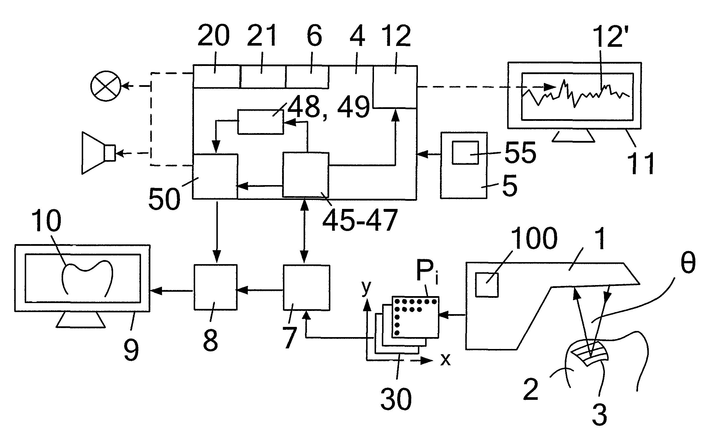
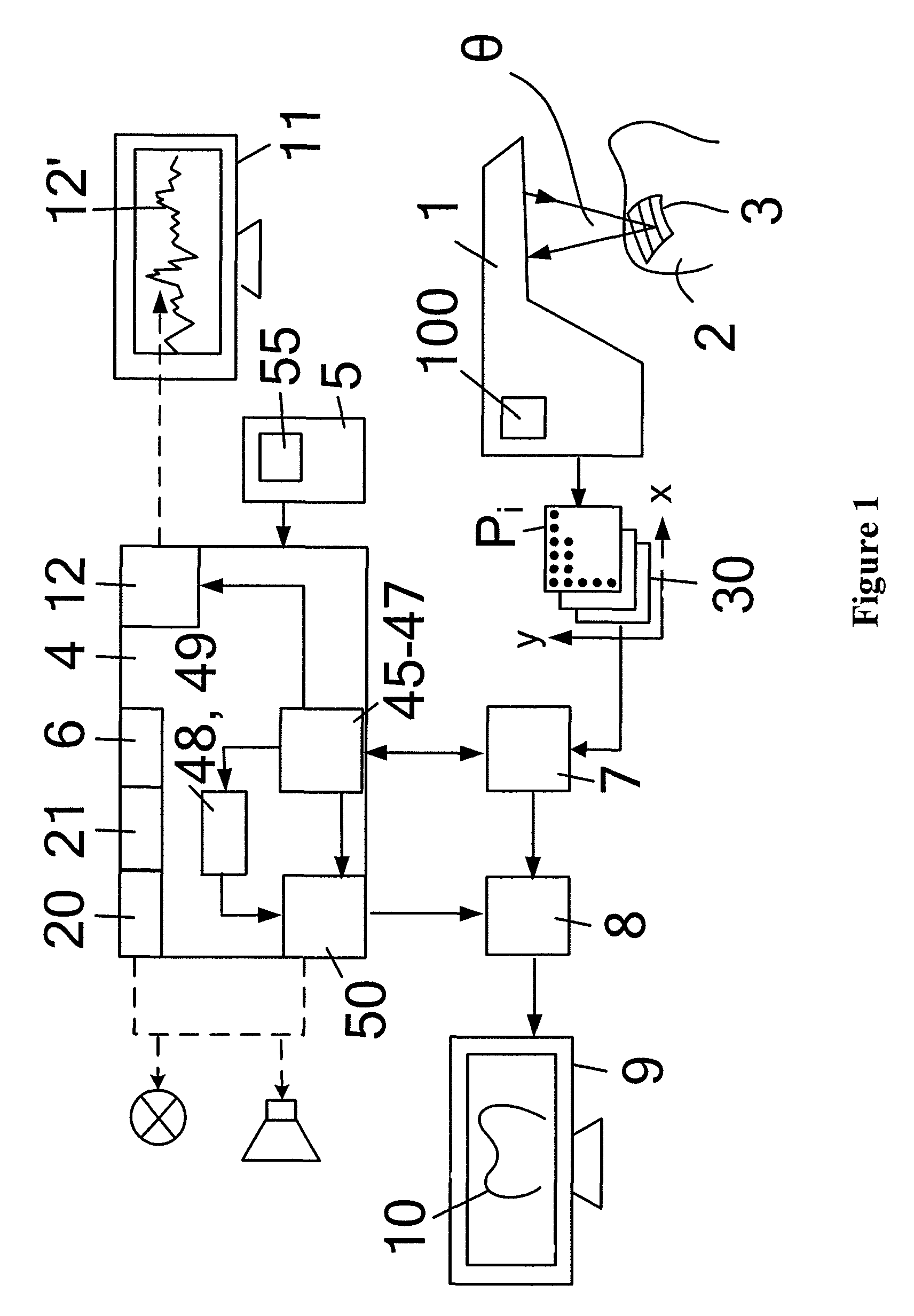
[0083]FIG. 1 is a diagram for clarification of the method of the invention and the device of the invention as exemplified by the phase shifting method. A dental 3D camera 1 is used in order to scan a three-dimensional object 2, namely a tooth, using a triangulation method. A viewfinder image, not shown but known from the prior art, makes it possible to position the 3D camera 1 over the object 2. The 3D camera 1 can then record five individual images 30 of the pattern 3 projected on the object 2, which pattern 3 can consist of parallel stripes having a sinusoidal brightness distribution, and the images 30 can each contain a plurality of pixels Pi having the coordinates (xi, yi). For each image 30 the pattern 3 can be shifted by a quarter of a period, i.e. a phase of 90°, the first four of the five images 30 forming a scanning sequence, from which a 3D data set 10 of the object 2 can be computed.

[0084]The recorded images 30 can be transmitted by the 3D camera 1 to an image memory 7 an...


PUM


Abstract

A dental 3D camera for optically scanning a three-dimensional object, and a method for operating a dental 3D camera. The camera operates in accordance with a triangulation procedure to acquire a plurality of images of the object. The method comprises forming at least one comparative signal based on at least two images of the object acquired by the camera while at least one pattern is projected on the object, and determining at least one camera shake index based on the at least one comparative signal.

Description

TECHNICAL FIELD[0001]The invention relates to a method and a device for optical scanning of three-dimensional objects by means of a dental 3D camera using a triangulation method. To this end, several individual images of a pattern projected on the object are recorded.PRIOR ART[0002]With prior methods, the user has had to fix the moment of initiation by actuating a footswitch and has had to hold the camera still from this moment on for the duration of the exposure. Recording was carried out at the moment of initiation and the result then visualized. When assessing the resulting image, a certain degree of experience was required for recognition of artifacts caused by camera shake and, if necessary, for a decision to erase the recording. The actuation of the footswitch is likely to increase movement at the moment of initiation. The problem of camera shake was thus solved by making a plurality of recordings Until one occurred in which, according to the judgment of the user, no artifacts...

Claims


Application Information

Patent Timeline
Patent Type & Authority Patents(United States)
IPC IPC(8): H04N13/02
CPCA61B5/1077A61B5/4547G01B11/2513A61C9/006G06T7/0057G01B11/2518G01B2210/52G01B11/2527G06T7/521
Inventor PFEIFFER, JOACHIMKLEIN, KONRAD
Owner SIRONA DENTAL SYSTEMS
Why Eureka
Try Eureka