Trichoplusia ni cell line and methods of use
a technology of trichoplusia ni and cell line, applied in the field of cell biology, medicine, agriculture, can solve the problems of insufficient quantity of membrane proteins, many other proteins are poorly expressed, and no gene expression system has been able to reliably express membrane proteins in sufficient quantities,
- Summary
- Abstract
- Description
- Claims
- Application Information
AI Technical Summary
Problems solved by technology
Method used
Image
Examples
example 1
Materials and Methods for Examples 2-5
[0072]Establishment of Cell Lines
[0073]T. ni eggs were obtained from Lloyd Browne, Entopath Inc., Easton, Pa. Embryonic tissues were prepared essentially as previously described (Lynn 1996, 2001). Briefly, eggs were surface sterilized with 0.05% sodium hypochlorite for five minutes, followed in succession by one five minute wash each in 70% ethanol, sterile distilled water, phosphate buffered saline (O'Reilly et al. 1992), and tissue culture medium. The embryos were then dissociated from the eggs into TC 100 (JRH Bioscience, Lenexa, Kans.) tissue culture medium, supplemented with 10% fetal bovine serum (Hyclone Laboratories), 100 U / ml penicillin, 100 μg / ml streptomycin, and 0.25 μg / ml amphotericin B. Pools of approximately 20 embryos were minced with a scalpel and transferred to disposable T12.5 flasks in 2 ml of medium supplemented as above. Cells were incubated at 27° C. Cells were monitored and fed every 7 days replacing half the media until ...
example 2
Cell Line Production
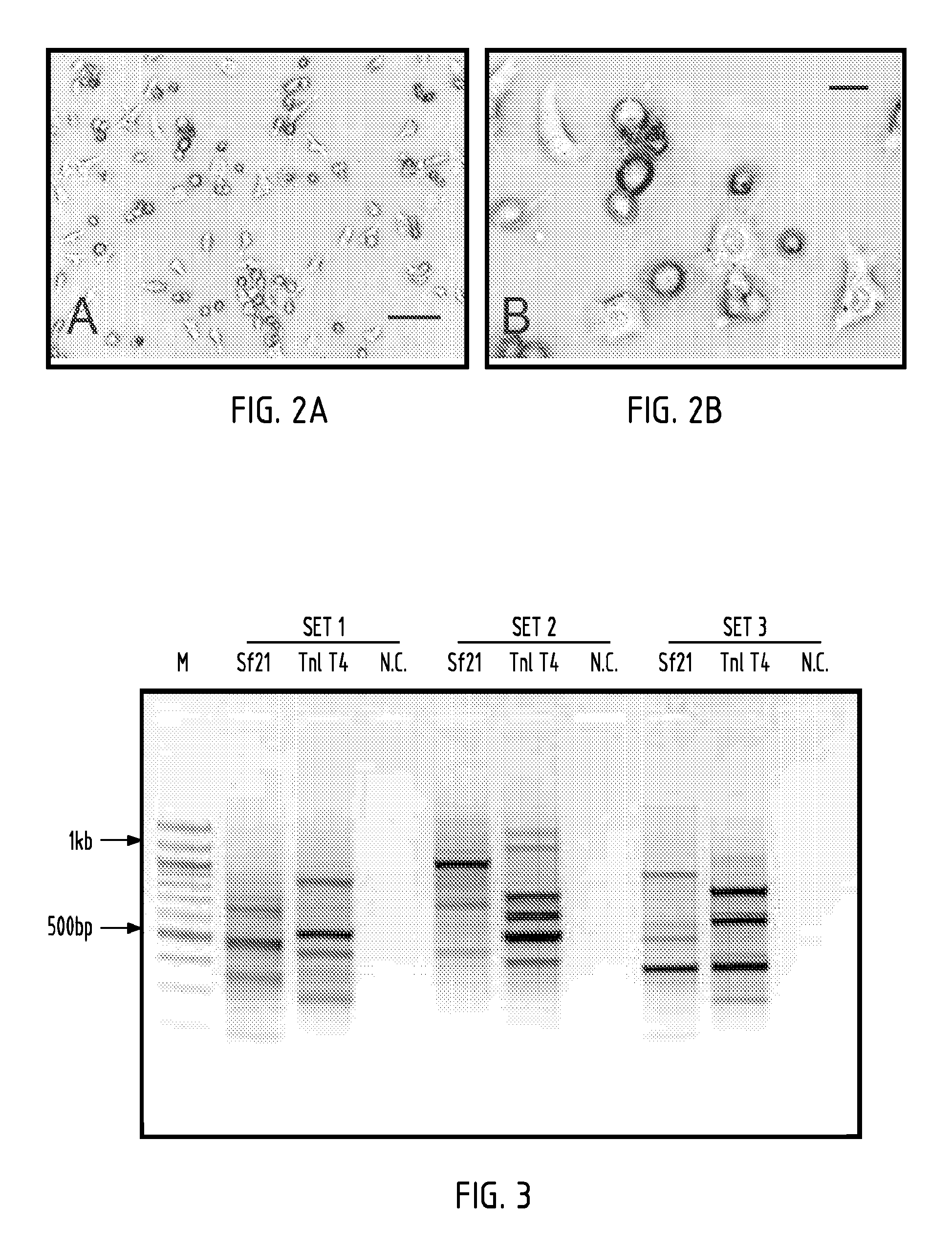
[0081]Cell lines were established from minced whole T. ni embryos in TC100 medium. TnT4 cells were obtained from one of eight flasks initiated in early 2005. These cells grew well and were split two months after initial seeding. Cells were split twice more at two-month intervals and subsequently at one-month intervals. One year after initiation, at passage number 15, cells were split once a week, at a split ratio of 1:4. Two years after initiation cells were subcultured twice a week at a split ratio of 1:14. TnT4 was selected for further characterization and development because it expressed the NTSR1-GFP-fusion protein, grew rapidly, and adapted readily to spinner culture. In plates, the TnT4 cells displayed variable morphologies including a small number of round cells and many cells with extended filipodia exhibiting diverse morphology (FIG. 2), but formed a contiguous sheet-like monolayer when confluent. When transferred to spinner culture the cells occasionall...
example 3
Cell Growth and Virus Production
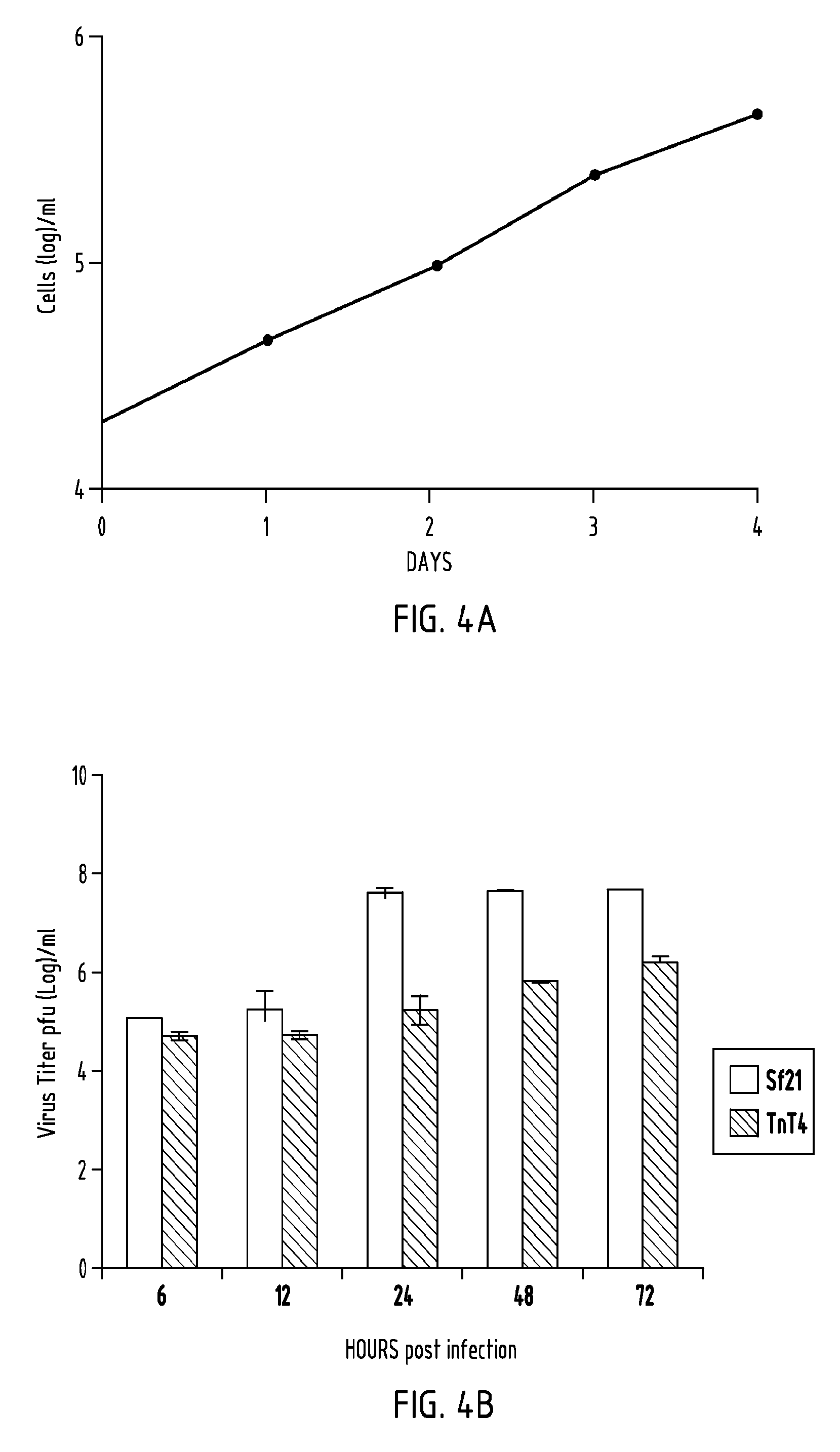
[0082]Cell growth rate was determined by counting cells, grown in tissue culture plates, at 24 h intervals (FIG. 4A). Calculated average cell doubling time was 21 hours. Production of AcMNPV budded virus in TnT4 cells was compared with that of Sf21 cells over time (FIG. 4B). TnT4 cells produced less BV than Sf21 cells over a 72 h period. There was also a delay in initial BV production in TnT4 cells compared to Sf21 cells (FIG. 4B, compare 12 and 24 h time points).
PUM
| Property | Measurement | Unit |
|---|---|---|
| Tm | aaaaa | aaaaa |
| temperature | aaaaa | aaaaa |
| temperature | aaaaa | aaaaa |
Abstract
Description
Claims
Application Information
 - R&D
- Intellectual Property
- Life Sciences
- Materials
- Tech Scout
- Unparalleled Data Quality
- Higher Quality Content
- 60% Fewer Hallucinations
Browse by: Latest US Patents, China's latest patents, Technical Efficacy Thesaurus, Application Domain, Technology Topic, Popular Technical Reports.
© 2025 PatSnap. All rights reserved.Legal|Privacy policy|Modern Slavery Act Transparency Statement|Sitemap|About US| Contact US: help@patsnap.com


