Anatomical model

an anatomical model and model technology, applied in the field of anatomical models, can solve the problems of many deficiencies of prior art anatomical models, difficult to pass through the anatomical model in a realistic manner, and not completely satisfactory,

Active Publication Date: 2013-03-26
OLYMPUS ENDO TECH AMERICA
View PDF32 Cites 59 Cited by
  • Summary
  • Abstract
  • Description
  • Claims
  • Application Information

AI Technical Summary

Benefits of technology

[0014]It is, therefore, an object of the present invention to provide an accurate and realistic anatomical model which can be used by physicians and other medical personnel to learn endoscopic insertion techniques and therapeutic procedures.
[0015]This and other objects are addressed by the provision and use of the present invention which, in one form of the invention, generally comprises an anatomical model comprising an inner lumen and an outer lumen, wherein the inner lumen is disposed inside of the outer lumen so as to create a space therebetween, and further wherein a fluid is disposed within the space, interior to the outer lumen and exterior to the inner lumen, whereby the inner lumen can accurately simulate the mucous membrane lining a mammalian tract (e.g., the gastrointestinal or genitourinary tract).

Problems solved by technology

However, there are many deficiencies associated with these prior art anatomical models.
Thus, forming the anatomical model out of an elastomer makes it difficult to pass the instrumentation (e.g., an endoscope) through the anatomical model in a realistic manner.
However, this approach is not completely satisfactory, since the lubricant can dry out, even in a relatively short time period, which can then make it even more difficult to pass instrumentation through the model.
Another significant deficiency of prior art anatomical models is that these models fail to realistically incorporate the external compression (e.g., abdominal pressure from adjacent organs) which acts on the gastrointestinal and / or genitourinary tract.
Prior art anatomical models are generally constructed with fixed supports which do not properly simulate the movable connections which can be characteristic of the gastrointestinal and / or genitourinary tracts.

Method used


Image

  • Anatomical model
  • Anatomical model
  • Anatomical model

Examples


Embodiment Construction

Anatomical Model Comprising an Inner Lumen and an Outer Lumen, and a Fluid Disposed in the Space Interior to the Outer Lumen and Exterior to the Inner Lumen

[0025]The present invention generally comprises an anatomical model comprising an inner lumen and an outer lumen, wherein the inner lumen is disposed inside of the outer lumen so as to create a space therebetween, and further wherein a fluid is disposed within the space, interior to the outer lumen and exterior to the inner lumen, whereby the inner lumen can accurately simulate the mucous membrane lining a mammalian tract (e.g., the gastrointestinal or genitourinary tract).

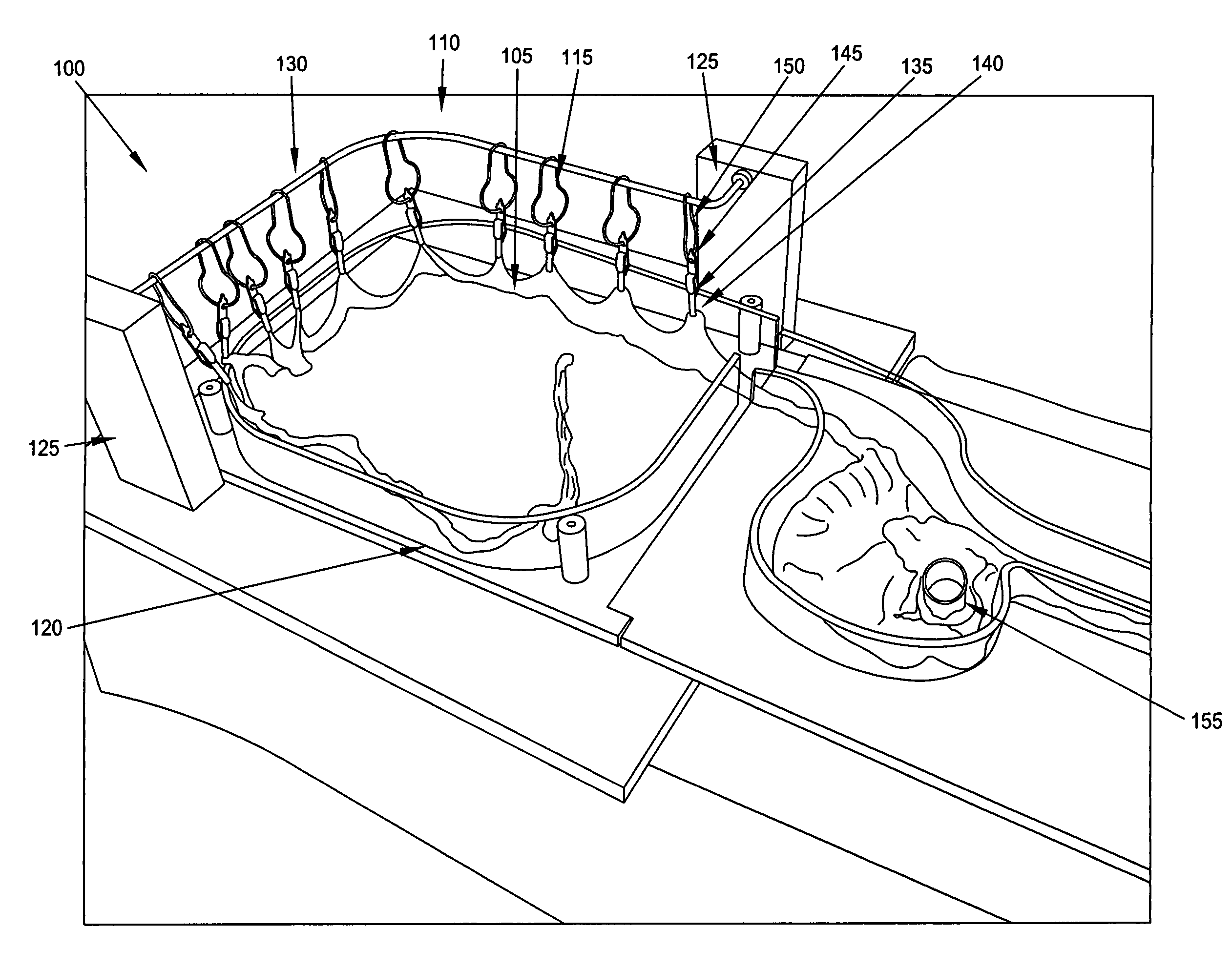
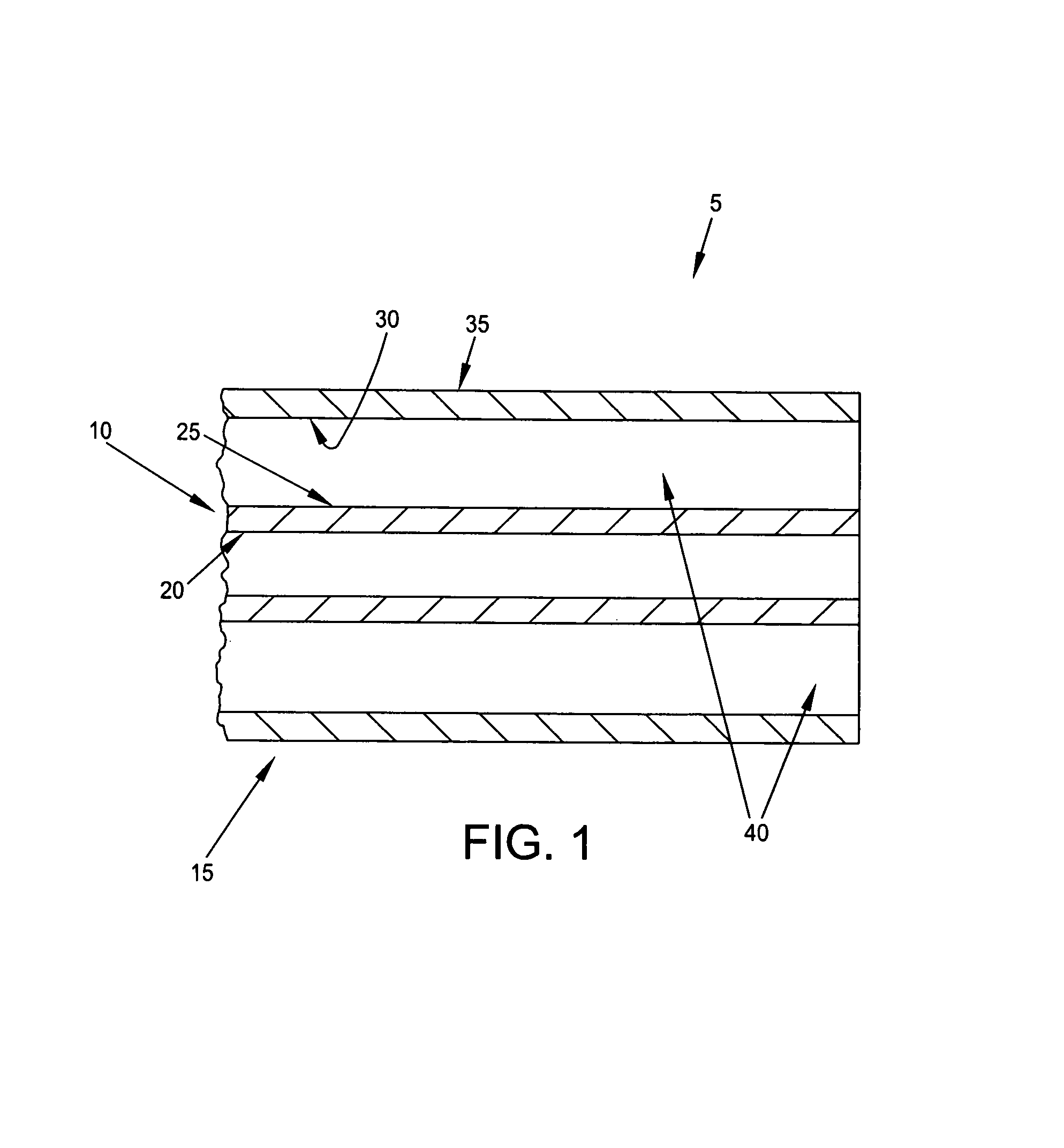
[0026]More particularly, and looking now at FIG. 1, there is shown an anatomical model 5 which comprises two lumens, an inner lumen 10 and an outer lumen 15, wherein inner lumen 10 is disposed inside of outer lumen 15. Inner lumen 10 generally comprises an interior surface 20 and an exterior surface 25. Outer lumen 15 generally comprises an interior surface 30 ...


PUM


Abstract

An anatomical model comprising:a lumen structure;a frame; andmovable connections selectively supporting the lumen structure to the frame.

Description

REFERENCE TO PENDING PRIOR PATENT APPLICATIONS[0001]This patent application:[0002](i) is a continuation-in-part of prior U.S. patent application Ser. No. 11 / 804,873, filed May 21, 2007 now U.S. Pat. No. 7,854,612 by James J. Frassica et al. for ANATOMICAL MODEL, which in turn claims benefit of prior U.S. Provisional Patent Application Ser. No. 60 / 801,719, filed May 19, 2006 by James Frassica et al. for ANATOMICAL MODEL PROV); and[0003](ii) claims benefit of prior U.S. Provisional Patent Application Ser. No. 61 / 000,539, filed Oct. 26, 2007 by James J. Frassica et al. for ANATOMICAL MODEL.[0004]The three above-identified patent applications are hereby incorporated herein by reference.FIELD OF THE INVENTION[0005]This invention relates to anatomical models in general, and more particularly to anatomical models of a mammalian tract for use in teaching endoscopic insertion techniques and therapeutic procedures to physicians and other medical personnel.BACKGROUND OF THE INVENTION[0006]In m...

Claims


Application Information

Patent Timeline
Patent Type & Authority Patents(United States)
IPC IPC(8): G09B23/28
CPCG09B23/285G09B23/30G09B23/32
Inventor FRASSICA, JAMES J.AILINGER, ROBERT E.AKERMAN, PAULBOOKWALTER, BILL
Owner OLYMPUS ENDO TECH AMERICA
Why Eureka
Try Eureka