Method for implanting prosthetic valve

a prosthetic valve and implanting technology, applied in the field of methods and devices for treating dysfunctional mitral valves, can solve the problems of congenital abnormalities or destruction of mitral valves, valves to be repaired or replaced, and may have severe physiological consequences

Active Publication Date: 2013-04-02
TRUELEAF MEDICAL LTD
View PDF11 Cites 245 Cited by
  • Summary
  • Abstract
  • Description
  • Claims
  • Application Information

AI Technical Summary

Benefits of technology

[0047]According to some embodiments of the invention, the valve is coated with a material which aids tissue growth.
[0049]In one embodiment of the invention, the expandable seating comprises self-expanding shoulders which assist in engaging surrounding leaflet tissue to secure the valve in position.

Problems solved by technology

Both of these valves may be damaged and dysfunctional, resulting in leakage during systole, requiring the valves to be repaired or replaced.
Mitral valve regurgitation causes heart murmurs and may have severe physiological consequences.
Occasionally, the mitral valve is congenitally abnormal or destroyed by infection or a bacterial endocarditis.
In the case of mitral stenosis, the valve orifice, i.e. the cross-section available for blood passage is reduced because of calcium nodes, leaflet thickening and / or reduced leaflet mobility, and, consequently, the valve does not allow normal blood flow.
The constant pressure overload of the left atrium may cause it to increase in size and become more prone to develop atrial fibrillation and to lose the atrial kick.
The loss of the atrial kick due to atrial fibrillation can cause a precipitous decrease in cardiac output.
A reduction in cardiac output, associated with acceleration of heart rate and shortening of the diastolic time, frequently leads to congestive heart failure.
As a result of the change of the left ventricular geometry, the posterior leaflet, which is attached to the posterior heart wall, is displaced and misaligned from the anterior leaflet which results in mitral regurgitation.
This weakens the leaflets and adjacent tissue, resulting in increased leaflet area and elongation of the chordae tendineae.
Elongation of the chordae tendineae often causes rupture.
The added workload creates an excessive strain on the left ventricle, which can lead to heart failure.
While patients with mild to moderate mitral regurgitation caused by MVP might experience no symptoms, increasing severity, even without symptoms, increases the load on the left ventricle.
Over time this can result in ventricular dilatation and congestive heart failure.
In some cases, such as when the valve is too damaged, mitral valves may require replacement.
However, they tend to degenerate over time and may require replacement within 10 to 15 years, necessitating a further operation.
Surgical valve replacement or repair is often a demanding operation as it requires cardiopulmonary bypass and it can expose patients, especially elderly ones, to many risks.
In transcatheter procedures, access to the native diseased valve is limited.
Such crimping was once considered detrimental to leaflet structure, causing tears and calcification to leaflets; however these issues have largely been resolved.
They are, however, generally expensive.
Replacement methods for aortic valves may be unsuitable for replacing mitral valves which differ therefrom, both anatomically and geometrically (morphologically).
Positioning and expanding a transcutaneous circular prosthetic valve (e.g. transcutaneous prosthetic aortic valve) within the opening of the ellipse shaped diseased mitral valve may result in inadequate sealing of the mitral valvular annulus, thereby resulting in severe regurgitation.
Furthermore, a circular valve deployed in a standard fashion will have poor anchoring and the valve is prone to detach and migrate from its position, to the detriment of the patient.
If the chordae are severed, then the ventricular wall is no longer anchored to the valve apparatus and the tethering effect of the chordae is lost.
As a result, left ventricular wall stress increases and left ventricular function deteriorates.
The main disadvantage of the methods for valve implantation described in the aforementioned patents is that they are not suitable for mitral valve replacement, mainly due to lack of or insufficient or cumbersome anchorage of the apparatus, and their application tends to risk continued regurgitation, i.e. blood backflow from left ventricle to left atrium upon systole.
Furthermore, the prosthetic valve apparatus is inserted over the native valve and thus there is increased risk of rupturing chordae tendineae or damaging the healthy leaflet.
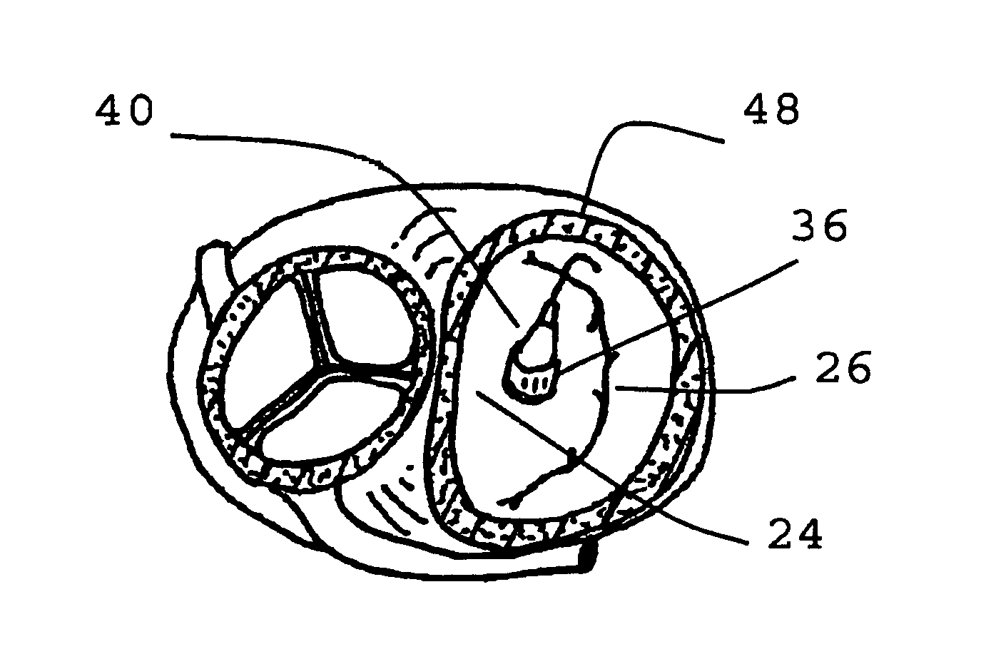
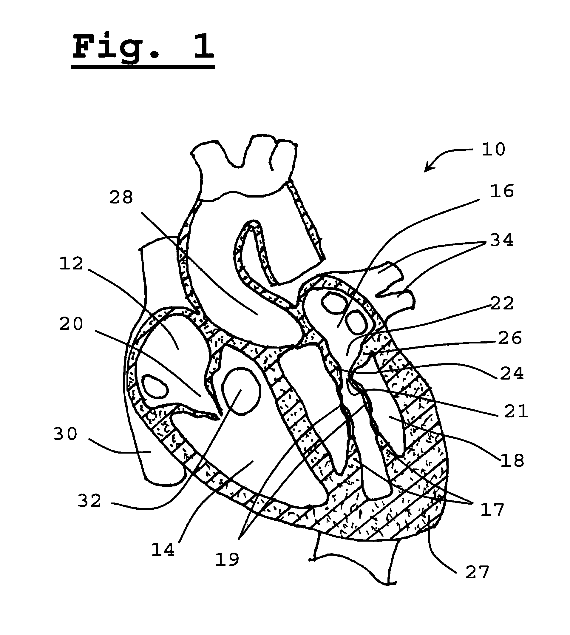
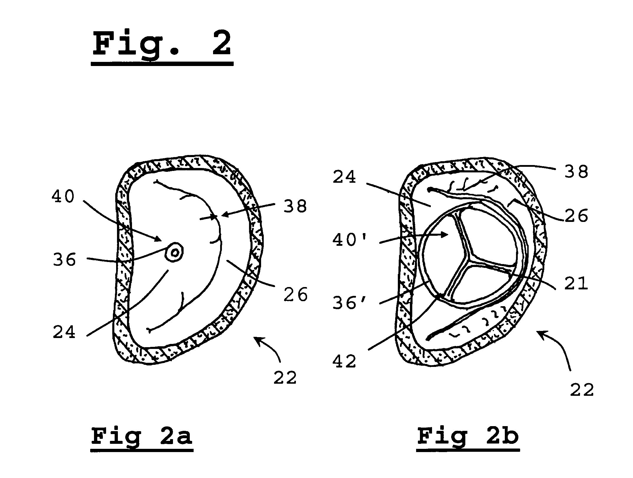
Method used


Image

  • Method for implanting prosthetic valve
  • Method for implanting prosthetic valve
  • Method for implanting prosthetic valve

Examples


Embodiment Construction

[0069]The present invention relates to a method for implanting a prosthetic valve apparatus in a novel and unexpected location. In this manner, the function of a prosthetic mitral valve is provided by the prosthetic valve and the native dysfunctional valve is voided, thereby avoiding paravalvular leakage. The method takes advantage of the special structural features of the tissue of the native leaflets and annulus of the mitral valve to keep the valve secured in place. The anterior leaflet extends across the annulus, which is an area of heart wall tissue at the junction of the atrial and ventricular walls that is relatively fibrous and significantly stronger than leaflet tissue. It provides structural support for the prosthetic valve and prevents its detachment from the novel orifice. It will be noted that the free edge of the valve leaflet has a reinforced, sinewy structure that is significantly stronger than central portions of the leaflet.

[0070]The approach of the invention is in...


PUM


Abstract

A method for implanting a prosthetic valve apparatus in a novel location which replaces the function of a native diseased valve. The prosthetic valve apparatus includes a one way valve and an expandable valve seating. anchoring and securing apparatus in a newly created orifice near or at the center of the anterior valve leaflet. The prosthetic valve apparatus also causes the sealing of the native valve and thus results in a solution for paravalvular leakage and regurgitation.

Description

FIELD OF THE INVENTION[0001]The present invention relates to methods and devices for treating dysfunctional mitral valves, particularly to new medical procedures for installing a prosthetic valve in a new location.BACKGROUND OF THE INVENTION[0002]Atrioventricular (AV) valves are cardiac valves that prevent backflow from the ventricles into the atria during systole. They are anchored to the wall of the heart at the fibrous skeleton by anchoring tendons named chordae tendineae. The chordae tendineae are attached to papillary muscles. Together, the papillary muscles and the chordae tendineae keep the valves from prolapsing into the atria when they close during systole. The actual opening and closing of the valves is caused by the pressure gradient across the valve. The left side AV valve is a bicuspid valve having two flaps or leaflets, and is commonly known as the mitral valve due to its shape being reminiscent of a bishop's mitre. The right side AV valve is a tricuspid valve, having ...

Claims


Application Information

Patent Timeline
Patent Type & Authority Patents(United States)
IPC IPC(8): A61B19/00A61F2/24
CPCA61F2/2463A61F2/2412A61F2/2433
Inventor SPENSER, BENJAMIN
Owner TRUELEAF MEDICAL LTD
Why Eureka
Try Eureka