
Method and system for spine visualization in 3D medical images

a 3d medical image and spine technology, applied in the field of spine visualization in 3d medical images, can solve the problems of difficult to see the curvature and abnormalities of the spine, difficult to obtain an unobstructed view of the spine using standard volume rendering techniques, and tedious task of volumetric data

Active Publication Date: 2013-04-16
SIEMENS HEALTHCARE GMBH
View PDF14 Cites 8 Cited by
  • Summary
  • Abstract
  • Description
  • Claims
  • Application Information

AI Technical Summary

Benefits of technology

The present invention is a way to automatically create 3D images of the spine that are easier to analyze and segment. It can improve the quality of the spine image and make it easier to see.

Problems solved by technology

However, using standard 2D slices to analyze spine information in the volumetric data is a tedious task, and it can be hard to see curvature and abnormalities of the spine since the spine passes through slices at an angle.
It is possible to use volume rendering to view the volumetric data in 3D, but it is difficult to obtain an unobstructed view of the spine using standard volume rendering techniques because other anatomic structures, such as the ribs, block the view of the spine.
The disadvantages of this method include the computational expense required from creating a distance function in the entire volume, a lack of robustness due to the specific choice of threshold, a high potential for overfitting due to the allowance of very high degree polynomials, and a lack of validation on the method itself with only one dataset tested.
The method is not fully automatic and requires user input in order to complete, limiting its use for clinical applications.
Finally, the method does not guarantee smooth transitions within the determined contour, limiting its utility in visualization.

Method used


Image

  • Method and system for spine visualization in 3D medical images
  • Method and system for spine visualization in 3D medical images
  • Method and system for spine visualization in 3D medical images

Examples


Embodiment Construction

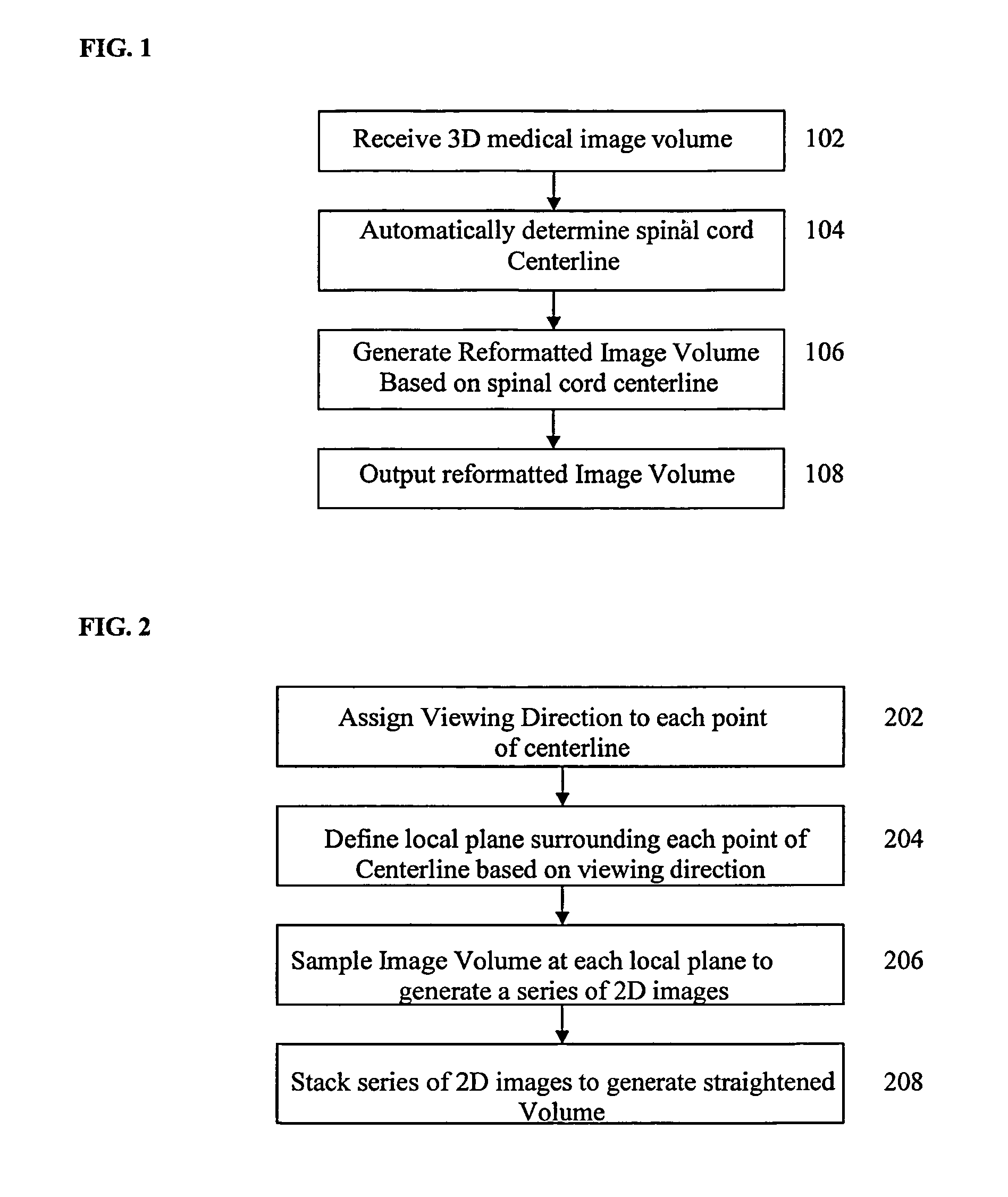
[0016]The present invention is directed to a method for visualizing the spine in 3D medical images. Embodiments of the present invention are described herein to give a visual understanding of the spine visualization method. A digital image is often composed of digital representations of one or more objects (or shapes). The digital representation of an object is often described herein in terms of identifying and manipulating the objects. Such manipulations are virtual manipulations accomplished in the memory or other circuitry / hardware of a computer system. Accordingly, is to be understood that embodiments of the present invention may be performed within a computer system using data stored within the computer system. For example, according to various embodiments of the present invention, electronic data representing a 3D medical image is manipulated within a computer system in order to reformat the image to visualize the spine.

[0017]FIG. 1 illustrates a method for visualizing the spi...


PUM


Abstract

A method and system for visualizing the spine in 3D medical images is disclosed. A spinal cord centerline is automatically determined in a 3D medical image volume, such as a CT volume. A reformatted image volume is then generated based on the spinal cord centerline. The reformatted image volume can be a straightened spine volume or a Multi-planar Reconstruction (MPR) based volume that follows the natural curve of the spine. The reconstructed volume can be displayed as 2D slices or 3D volume renderings.

Description

[0001]This application claims the benefit of U.S. Provisional Application No. 60 / 938,727, filed May 18, 2007, the disclosure of which is herein incorporated by reference.BACKGROUND OF THE INVENTION[0002]The present invention relates spine visualization in 3D medical images, and more particularly to reformatting 3D medical images for improved visualization of the spine.[0003]3D medical images, or volumetric medical data, such as computed tomography (CT) images, typically contain a wealth of anatomical information. For example, a chest CT image taken to evaluate a patient's airways may also contain valuable information concerning the patient's spine. In order to view the spine in such CT image, the volumetric data can be analyzed using standard two 2D slices. However, using standard 2D slices to analyze spine information in the volumetric data is a tedious task, and it can be hard to see curvature and abnormalities of the spine since the spine passes through slices at an angle. It is ...

Claims


Application Information

Patent Timeline
Patent Type & Authority Patents(United States)
IPC IPC(8): A61B5/05
CPCA61B5/103A61B5/4561G06T7/0085G06T7/0091G06T7/60G06T15/08A61B5/055A61B8/00G06K2209/055G06T2207/20044G06T2207/30012G06T2207/30172G06T2215/06A61B5/407G06T7/13G06T7/155G06V2201/033
Inventor KIRALY, ATILLA PETERALVINO, CHRISTOPHER V.SHEN, HONG
Owner SIEMENS HEALTHCARE GMBH
Learn More