Magnetic resonance imaging apparatus and method configured for susceptibility-emphasized imaging with improved signal-to-noise ratio

a magnetic resonance imaging and signal-to-noise ratio technology, applied in the field of susceptibility-emphasized imaging, can solve the problems of long imaging time, inability to be set short, and the signal-to-noise ratio deteriorates when an image is taken, and achieves the effect of preferable signal-to-noise ratio and susceptibility-emphasized

Active Publication Date: 2013-06-18
FUJIFILM HEALTHCARE CORP
View PDF13 Cites 2 Cited by
  • Summary
  • Abstract
  • Description
  • Claims
  • Application Information

AI Technical Summary

Benefits of technology

[0017]Thus, it is possible to obtain a susceptibility-emphasized image having a preferable signal-to-noise ratio in the susceptibility-emphasized imaging using the echo planar method in the MRI apparatus.

Problems solved by technology

As a result, repetition time (TR) of a pulse sequence (hereinafter simply abbreviated as sequence) can not be set short, whereby taking a long period of time for imaging.
In the case that the echo planar method is applied to susceptibility-emphasized imaging, since the echo signal having a long echo time tends to be greatly attenuated, the unsolved problem remains that the signal-to-noise ratio deteriorates when an image is reconstructed from such echo signals.

Method used


Image

  • Magnetic resonance imaging apparatus and method configured for susceptibility-emphasized imaging with improved signal-to-noise ratio
  • Magnetic resonance imaging apparatus and method configured for susceptibility-emphasized imaging with improved signal-to-noise ratio
  • Magnetic resonance imaging apparatus and method configured for susceptibility-emphasized imaging with improved signal-to-noise ratio

Examples


first embodiment

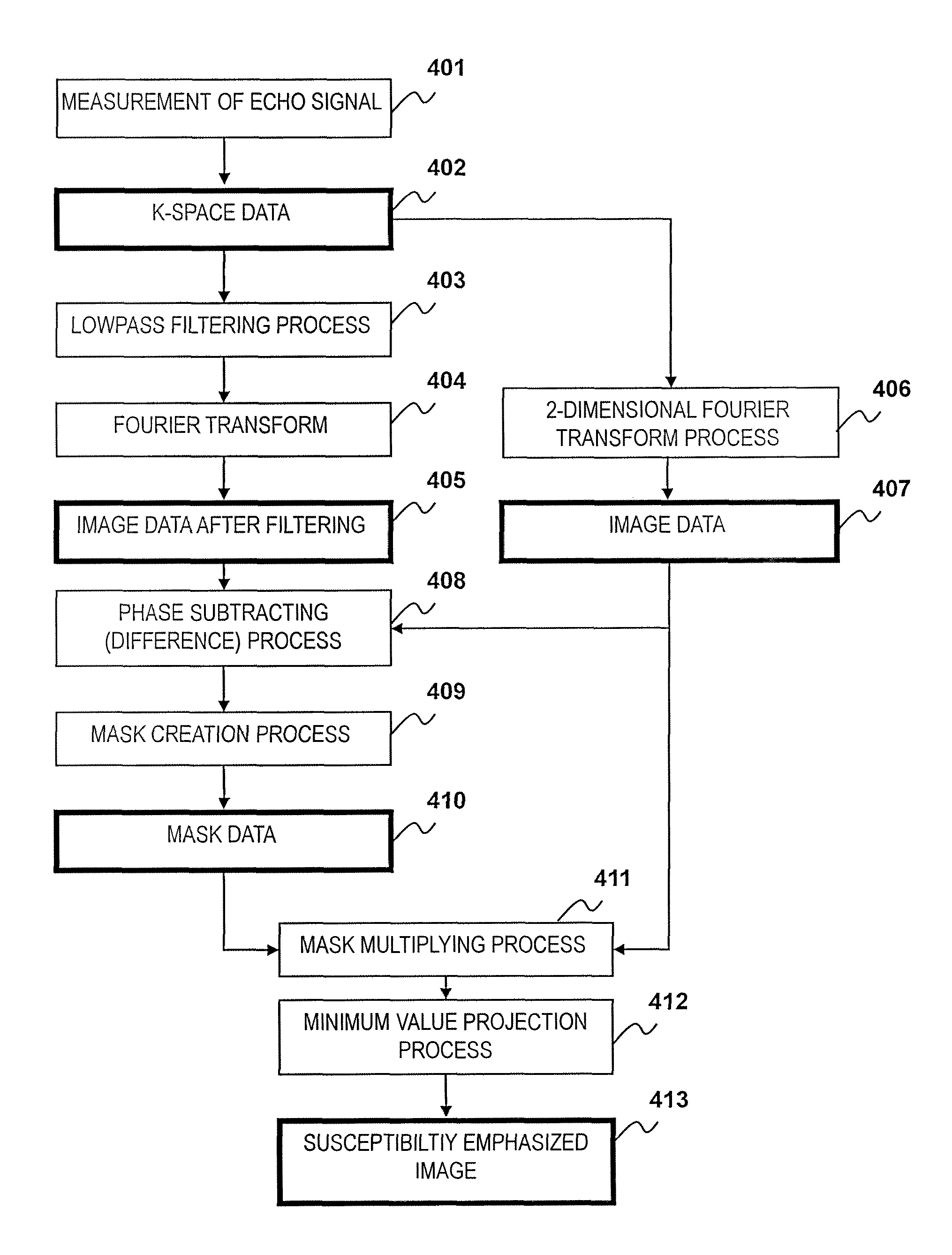
[0066]The first embodiment of the MRI apparatus and the susceptibility-emphasized imaging method related to the present invention will be described. The present embodiment divides the echo signal group measured using the echo planar method into a first echo signal group measured in the first half and a second echo signal group measured in the last half, and creates image data from the first echo signal group and mask data from the second echo signal group. The embodiment will be described in detail based on FIG. 5 and FIG. 6.

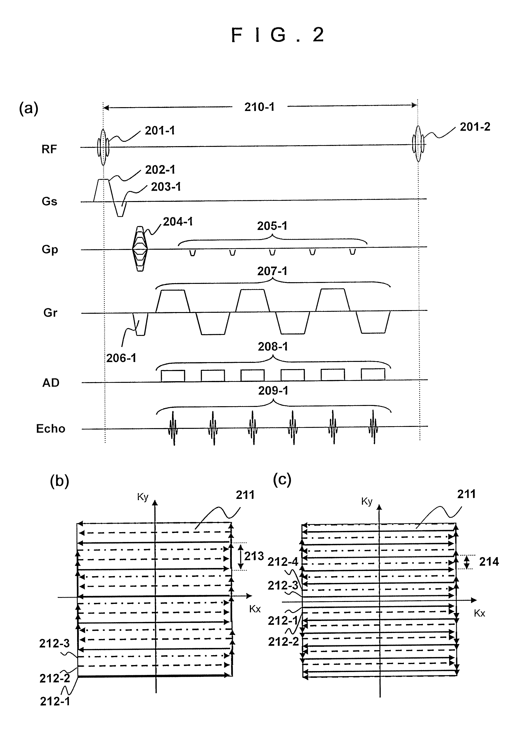
[0067]First, the sequence of the multi-shot echo planar method of the gradient echo type related to the present embodiment and the K-space data acquired using the sequence will be described referring to FIG. 5. FIG. 5(a) shows only phase encode gradient magnetic field axis (Gp) and an echo signal from among the sequences described in FIG. 2(a), and the other sequences are omitted since they are the same as FIG. 2(a). While an example using the echo planar method...

second embodiment

[0093]Next, the second embodiment of the MRI apparatus and the susceptibility-emphasized imaging method related to the present invention will be described. The present embodiment applies a phase blip gradient magnetic field pulse for every two echo signals, and the echo signal group measured in odd-numbered order is set as a first echo signal group and the echo signal group measured in even-numbered order is set as a second echo signal group. The differences from the first embodiment are that the sequence diagram which applies a phase blip gradient magnetic field pulse for every 2 echo signals, and the placement of data in the K-space. The present embodiment will be described below in detail by explaining only the difference from the first embodiment, referring to FIG. 9.

[0094]First, the sequence diagram of the present embodiment will be described based on FIG. 9(a). FIG. 9(a) is the sequence diagram of the multi-shot echo planar method of the gradient echo type related to the prese...

third embodiment

[0105]Next, the third embodiment of the MRI apparatus and the susceptibility-emphasized imaging related to the present invention will be described. The present embodiment reduces imaging time by making the number of measurement of echo signals for a mask and the number of measurement of echo signals for an image different. The differences from the first embodiment are the sequence diagram due to the difference of the measurement numbers of echo signals for a mask and echo signals for an image, and the placement of data in the K-space. Only the difference from the first embodiment will be described below.

[0106]First, the sequence diagram of the present embodiment will be described using FIG. 10(a). FIG. 10(a) shows the sequence diagram by the multi-shot echo planar method of the gradient echo type related to the present embodiment, and only an echo signal (Echo) measured by one repetition (shot). Explanation on the other parts will be omitted since they are the same as the sequence i...


PUM


Abstract

A susceptibility-emphasized image having a preferable signal-to-noise ratio can be obtained by a method including measuring a plurality of echo signals using the echo planar method, dividing the plurality of echo signals into a first echo signal group and a second echo signal group, acquiring an image data from the first echo signal group while acquiring a mask data from the second echo signal group, and obtaining the susceptibility-emphasized image through use of the image data and the mask data.

Description

TECHNICAL FIELD[0001]The present invention is related to a technique for acquiring a susceptibility-emphasized image with high speed using a magnetic resonance imaging (hereinafter referred to as MRI) apparatus that acquires a tomographic image of an examination region of an object to be examined utilizing nuclear magnetic resonance phenomenon.BACKGROUND ART[0002]An MRI apparatus uses a uniform static magnetic field, and the static magnetic field thereof changes locally depending on the magnetic susceptibility of an object. The effect of the local magnetic field change appears in image data as change of a phase. The imaging method that emphasizes the phase change by arithmetic processing is known as susceptibility-emphasized imaging (Patent Document 1). Susceptibility-emphasized imaging has received attention as a method effective for MR angiography of a venous blood vessel, since it is capable of emphasizing magnetic susceptibility due to deox hemoglobin in the blood.[0003]However,...

Claims


Application Information

Patent Timeline
Patent Type & Authority Patents(United States)
IPC IPC(8): G01V3/00
CPCG01R33/5616G01R33/5635G01R33/5617
Inventor TAKIZAWA, MASAHIRO
Owner FUJIFILM HEALTHCARE CORP
Why Eureka
Try Eureka