Marker for identification of tissue type of epithelial ovarian cancer, and method for determination of the occurrence of epithelial ovarian cancer based on tissue type by using the marker

a tissue type and marker technology, applied in the field of molecular typing, can solve the problems of not being used in early diagnosis, low positive rate of markers, and inability to establish a definitive diagnosis method, etc., and achieve the effect of high specificity and high reliability

Active Publication Date: 2013-08-27
SHIMADZU CORP
View PDF4 Cites 2 Cited by
  • Summary
  • Abstract
  • Description
  • Claims
  • Application Information

AI Technical Summary

Benefits of technology

[0022]Therefore, it was also found that more accurate identification of a tissue-type can be realized by using not only the above tumor marker singly, but also using an appropriate combination of the protein that is specifically significantly expressed in two kinds of tissue-types and the protein whose expression is downregulated in a specific one kind of tissue-type as markers for identification of a tissue-type.
[0235]According to the present invention, it is possible to identify the morbidity of epithelial ovarian cancer based on a tissue-type in view of molecular typing which is different from a conventional histopathology. In other words, according to the present invention, it is possible to provide a marker for identification of a tissue-type of epithelial ovarian cancer.
[0236]The marker of the present invention was found by profiling based on identification of a protein. While profiling is generally executed by immunostaining, the profiling executed by the present inventors realizes high reliability as a marker of the found protein unlike that obtained by immunostaining where artifacts are often observed.
[0237]Furthermore, since markers of the present invention are found based on quantification of a difference in expression between tissue-types, they are clinically very persuasive. Furthermore, by using a combination of a plurality of markers, it is possible to execute analysis with higher specificity than in the case where a conventionally used broad marker having lower specificity (for example, CA-125 or CA-19-9) is used.

Problems solved by technology

Therefore, early diagnosis would be the most important issue for improving the long term survival rate.
However, most of ovarian cancers in early stage are silent, and a noninvasive definitive diagnosis method has not been established.
On the contrary, however, the marker shows a low positive rate and too poor specificity in early stage ovarian cancer.
Therefore, it has a drawback that it can not be used in early diagnosis.
However, as described in Nature, 2004, there is an opinion that the methodology lacks a scientific basis.
For this reason, this method has not been brought into practical use yet.

Method used


Image

  • Marker for identification of tissue type of epithelial ovarian cancer, and method for determination of the occurrence of epithelial ovarian cancer based on tissue type by using the marker
  • Marker for identification of tissue type of epithelial ovarian cancer, and method for determination of the occurrence of epithelial ovarian cancer based on tissue type by using the marker
  • Marker for identification of tissue type of epithelial ovarian cancer, and method for determination of the occurrence of epithelial ovarian cancer based on tissue type by using the marker

Examples


example 1

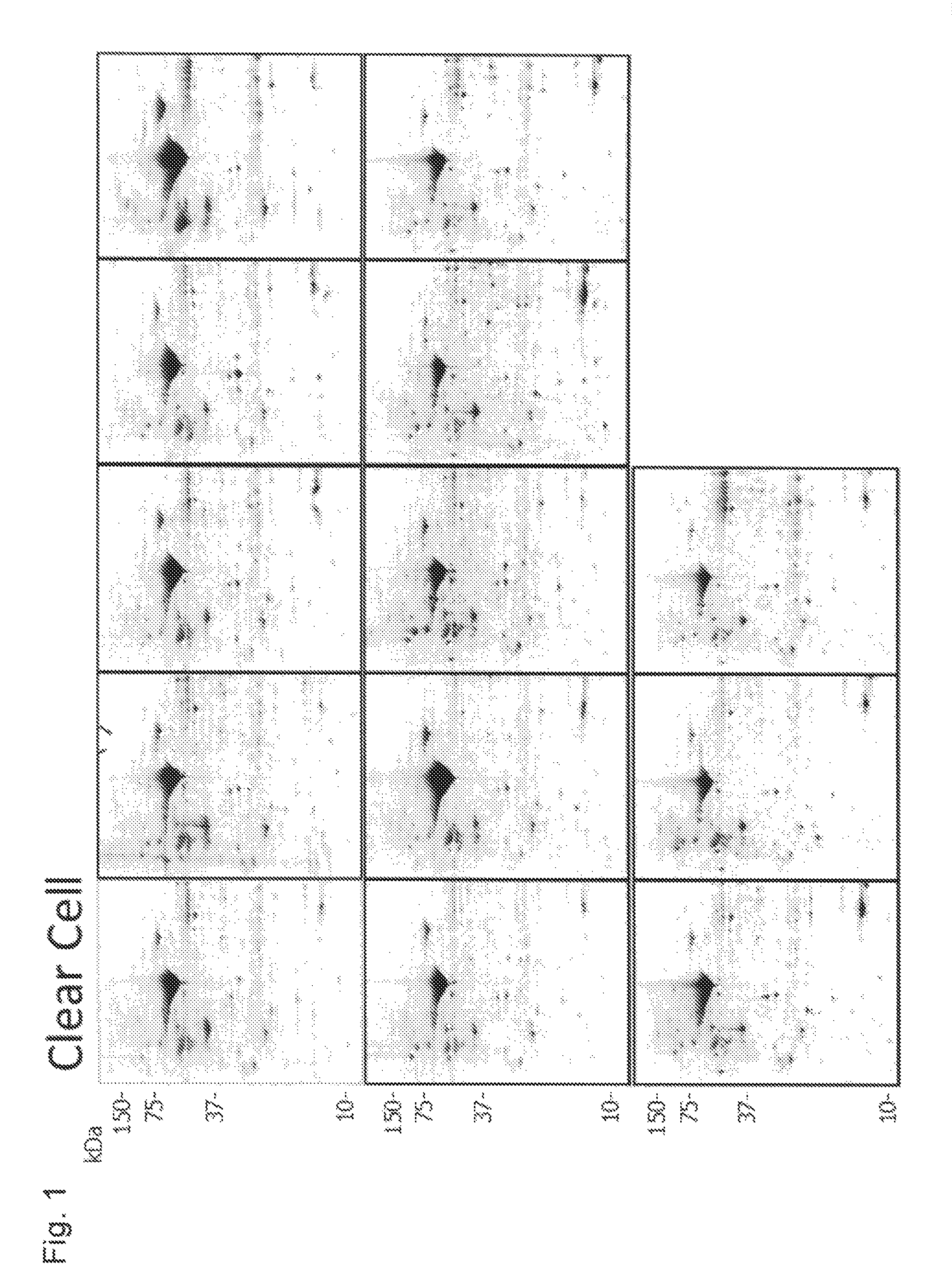
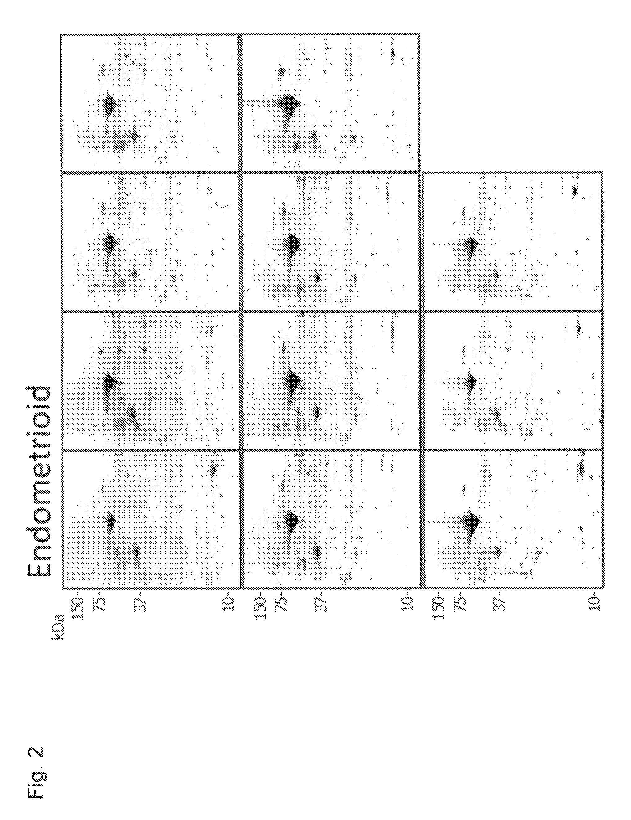
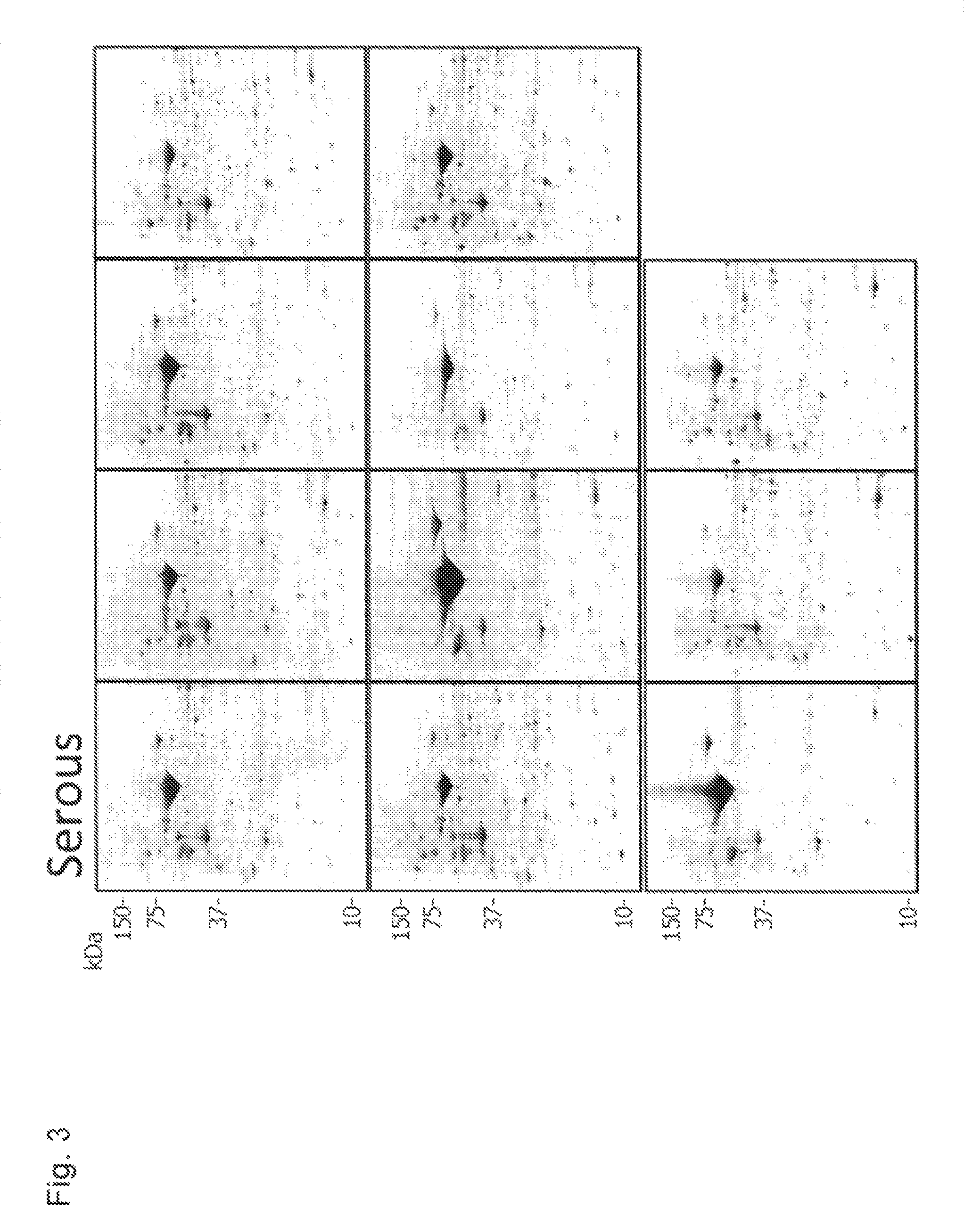
[0407]Profiling was conducted using pathological tissues of epithelial ovarian cancer that had been excised from patients, and for which accurate tissue-type diagnosis had been made from the view point of histopathology (4 tissue-types and 41 specimens: clear cell cancer 13 specimens, endometrioid carcinoma 11 specimens, serous adenocarcinoma 11 specimens, and mucinous adenocarcinoma 6 specimens).

[0408]An excised pathological tissue (stocked in liquid nitrogen) was crushed into fragments of an appropriate size in the presence of liquid nitrogen. The crushed tissue specimen was transferred into a micro tube with a screw cap, and suspended in about 5 times weight of Lysis buffer (50 mM HEPES-NaOH pH 7.5, 100 mM NaCl, 2% CHAPS, 1% Triton X-100). The concentration in the state of a suspension was quantified by the Bradford method, and a specimen corresponding to 300 μg of protein was subjected to desalination by trichloroacetic acid precipitation, to prepare a sample for two-dimensional...

example 2

[0425]For screening the above-described 80 candidate markers into molecules having a determinative role in diagnosing a tissue-type of an unknown ovarian cancer tissue using a molecular technique, a classification prediction model using a decision tree was constructed using statistical analysis software SPSS (SPSS Inc.). Four tissue-types were used as dependent variables, and quantified values of 80 spots of candidate markers scaled into three stages (low expression, high expression, extra-high expression) were used as independent variables. The reference value in scaling was appropriately determined based on the expression pattern and the average quantified value of each candidate marker determined by profiling.

[0426]As one example, FIG. 11 shows a decision tree model for determining whether an unknown specimen is Clear Cell, prepared by using 41 specimens from which the profile was prepared. In FIG. 11, a protein used as a variable of divergence is shown inside the dashed line, an...

example 3

[0429]Using tissue extracts (excluding one specimen of mucinous adenocarcinoma) that are the same with those subjected to two-dimensional electrophoresis in Example 1, quantitative detection by Western blotting was conducted. As antibodies, anti-Annexin-A4 (D-2, Santa Cruz Biotechnology), anti-Maspin precursor (G167-70, BD Transduction Laboratories), anti-Cellular retinoic acid-binding protein 2 (Bethyl Laboratories) and anti-Phosphoserine aminotransferase (Proteintech Group Inc.) were used.

[0430]Results of Western blotting by 40 specimens and 4 antibodies are shown in FIG. 12 (for Clear Cell and Endometrioid) and in FIG. 13 (for Serous and Mucinous). As shown in these drawings, a specific band was detected in a position of respective target molecular weight, and an expression pattern that seems to be tissue-specific was observed similarly to the result obtained by two-dimensional electrophoresis. Each band was quantified based on the band of β-actin (Beta actin) which was a loading...


PUM

PropertyMeasurementUnit
temperatureaaaaaaaaaa
thickaaaaaaaaaa
thickaaaaaaaaaa

Abstract

It is provided a method for identification of the morbidity of epithelial ovarian cancer based on a tissue-type in view of molecular typing which is different from a conventional histopathology, and a marker for identification of a tissue-type of epithelial ovarian cancer. A method for identification of the morbidity of epithelial ovarian cancer based on a tissue-type, comprising: subjecting a sample originated from an individual of interest to a treatment for detecting at least one selected from the group consisting of biological molecules specifically showing an upregulation in expression in a specific tissue-type of epithelial ovarian cancer, and / or at least one selected from the group consisting of biological molecules specifically showing a downregulation in expression in a specific tissue-type of epithelial ovarian cancer, and identifying whether or not the significant detection of the protein is achieved, thereby identifying the tissue-type.

Description

TECHNICAL FIELD[0001]The present invention relates to techniques of screening, diagnosis, examination, follow up, and classification of epithelial ovarian cancer (EOC). More specifically, the present invention relates to molecular typing of a tissue-type of epithelial ovarian cancer. In other words, the present invention relates to a marker for identification of a tissue-type of epithelial ovarian cancer and a method for identification of epithelial ovarian cancer based on a tissue-type using the same.BACKGROUND ART[0002]Ovarian cancer is a case whose number is the second highest in malignant tumors of female genitalia, and has the highest lethality. This mostly occurs in premenopausal or postmenopausal women. Only in the U.S., annually 26,000 people are newly diagnosed to suffer from ovarian cancer, and 16,000 patients are deceased (in 2004). Five year survival rates for classified stages are 90% for stage I, 70% for stage II, 25% for stage III, and 10% for stage IV, and prognosis ...

Claims


Application Information

Patent Timeline
Patent Type & Authority Patents(United States)
IPC IPC(8): G01N33/53
CPCG01N33/57449G01N2800/56
Inventor SATO, TAKA-AKITOYAMA, ATSUHIKOSHIMADA, TAKASHISUGITA, TETSUYOSHIAOKI, DAISUKESUZUKI, ATSUSHISUSUMU, NOBUYUKINOMURA, HIROYUKI
Owner SHIMADZU CORP
Why Eureka
Try Eureka