Method for performing micro-perimetry exams based on a retinal volume image and a well registered fundus image
a micro-perimetry and fundus image technology, applied in the field of ophthalmic diagnostic testing, can solve the problems of extremely limited techniques and achieve the effect of high detail
- Summary
- Abstract
- Description
- Claims
- Application Information
AI Technical Summary
Benefits of technology
Problems solved by technology
Method used
Image

Examples
Embodiment Construction
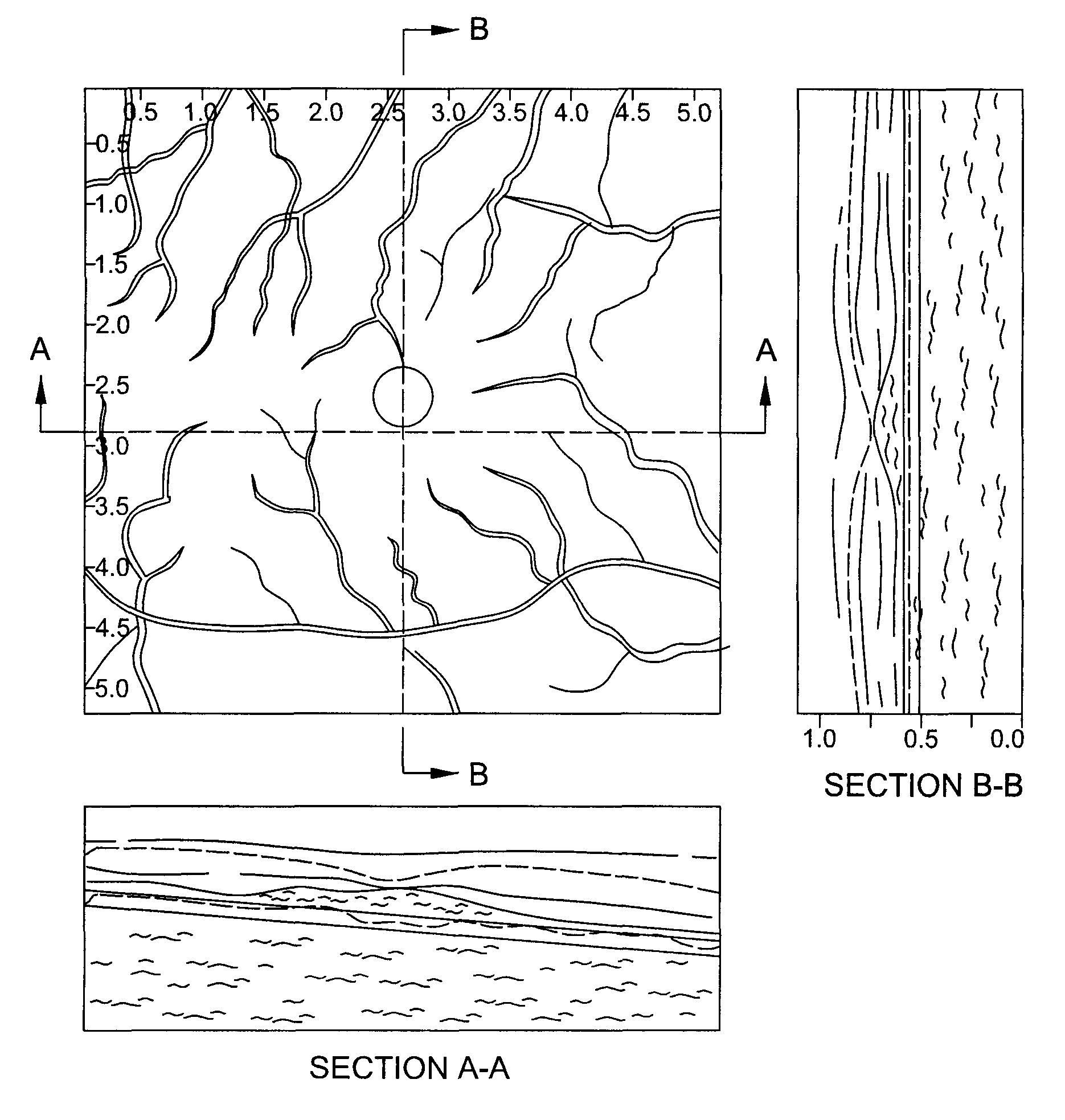
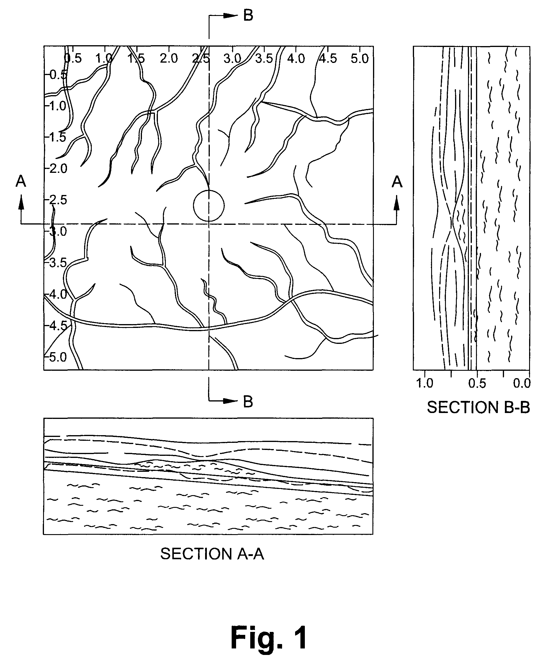
[0011]FIG. 1 shows an en-face image of the fundus of an eye, with sectional images obtained by OCT shown beside and below the en-face image. The image to the right is a cross section taken along the vertical axis, and the image below is a cross section taken along the horizontal axis. Such an image can be displayed on the display screen of OCT equipment.
[0012]In the case of SLO / OCT apparatus, the en-face image is obtained using SLO (scanning laser ophthalmoscope), whereas the sectional images are obtained by OCT.
[0013]In operation, the operator looks at the sectional OCT images to the right and below the main image to find points of interest, for example, particular points where some pathological condition may be noted. He or she then selects these points, for example, by pointing to them and clicking with a mouse. The computer forming part of the OCT equipment then automatically places marks (indicated by an X) at these points in the sectional images, and makes corresponding points...
PUM
 Abstract
Description
Claims
Application Information
 - R&D Engineer
- R&D Manager
- IP Professional
- Industry Leading Data Capabilities
- Powerful AI technology
- Patent DNA Extraction
Browse by: Latest US Patents, China's latest patents, Technical Efficacy Thesaurus, Application Domain, Technology Topic.
© 2024 PatSnap. All rights reserved.Legal|Privacy policy|Modern Slavery Act Transparency Statement|Sitemap