
Classification of medical diagnostic images

a medical diagnostic and image technology, applied in image data processing, character and pattern recognition, instruments, etc., can solve the problems of subjective visual assessment, time-consuming, and difficult visual assessment of disease severity and progression

Inactive Publication Date: 2014-08-19
UNIVERSITY OF COPENHAGEN
View PDF2 Cites 10 Cited by
  • Summary
  • Abstract
  • Description
  • Claims
  • Application Information

AI Technical Summary

Benefits of technology

The invention is about an automatic way to categorize medical images of lungs based on their likelihood of being related to lung diseases such as chronic obstructive pulmonary disease (COPD), cystic fibrosis, or severe asthma, or based on their severity. This approach uses supervised learning, without needing manually annotated data, and instead relies on pulmonary function testing (PFT) data or other metadata to identify the labels. Overall, the invention provides a fast, accurate, and automated way to analyze lung images and improve the diagnosis and treatment of lung diseases.

Problems solved by technology

Current quantitative measures of chronic obstructive pulmonary disease (COPD) are limited in several ways.
These are cheap and fast to acquire but are limited by insensitivity to early stages of COPD and lack of reproducibility [8].
However, it is difficult to assess disease severity and progression visually.
Moreover, visual assessment is subjective, time-consuming, and suffers from intra-observer and inter-observer variability [3, 18].
These measures consider each parenchyma voxel in the CT image independently thereby disregarding potentially valuable information, such as spatial relations between voxels of similar or differing intensities and patterns at larger scales, and they are restricted to a single threshold parameter, which makes them sensitive to noise in the CT images.
COPD is estimated to become the fifth most burdening disease and the third leading cause of death worldwide by 2020[23], and the limitations of current quantitative measures may hinder progression in research on treatments for the disease.
Manual annotation suffers from the same limitations as visual assessment of emphysema in CT images [3, 18], moreover, it is hard to demarcate or position ROIs in CT images, since the appearance of the disease patterns is subtle and diffuse, especially at early stages of COPD.
Further, analysis is limited to current knowledge and experience of the experts, and there can be a bias towards typical cases in the annotated data set.

Method used


Image

  • Classification of medical diagnostic images
  • Classification of medical diagnostic images
  • Classification of medical diagnostic images

Examples


Embodiment Construction

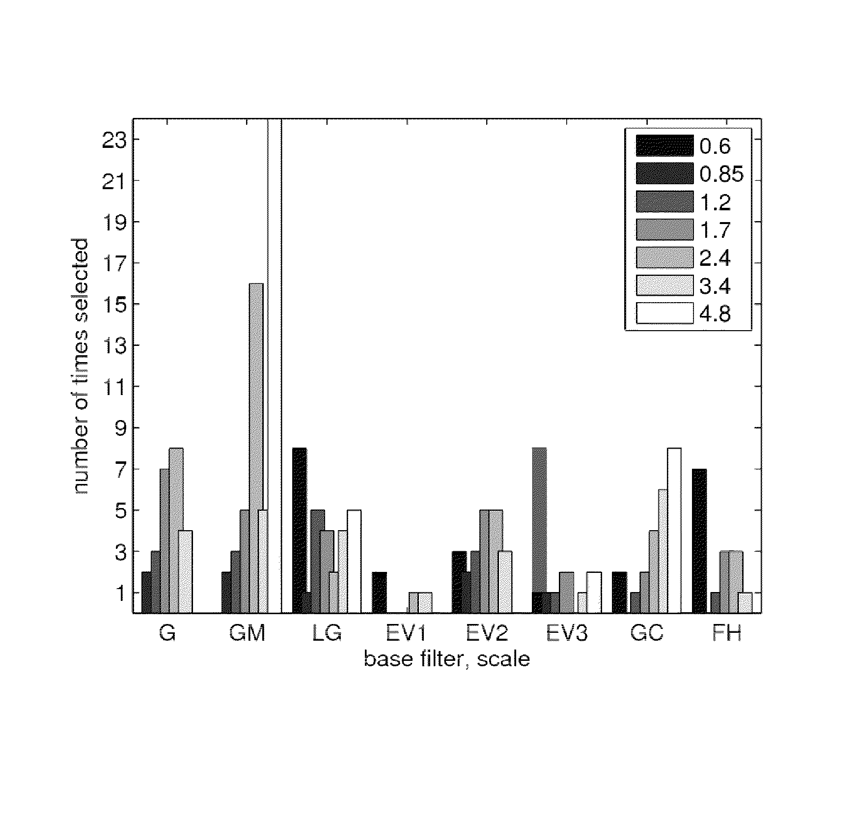
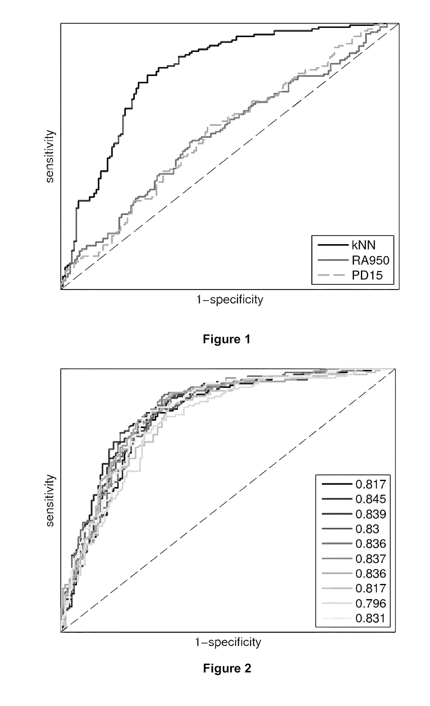
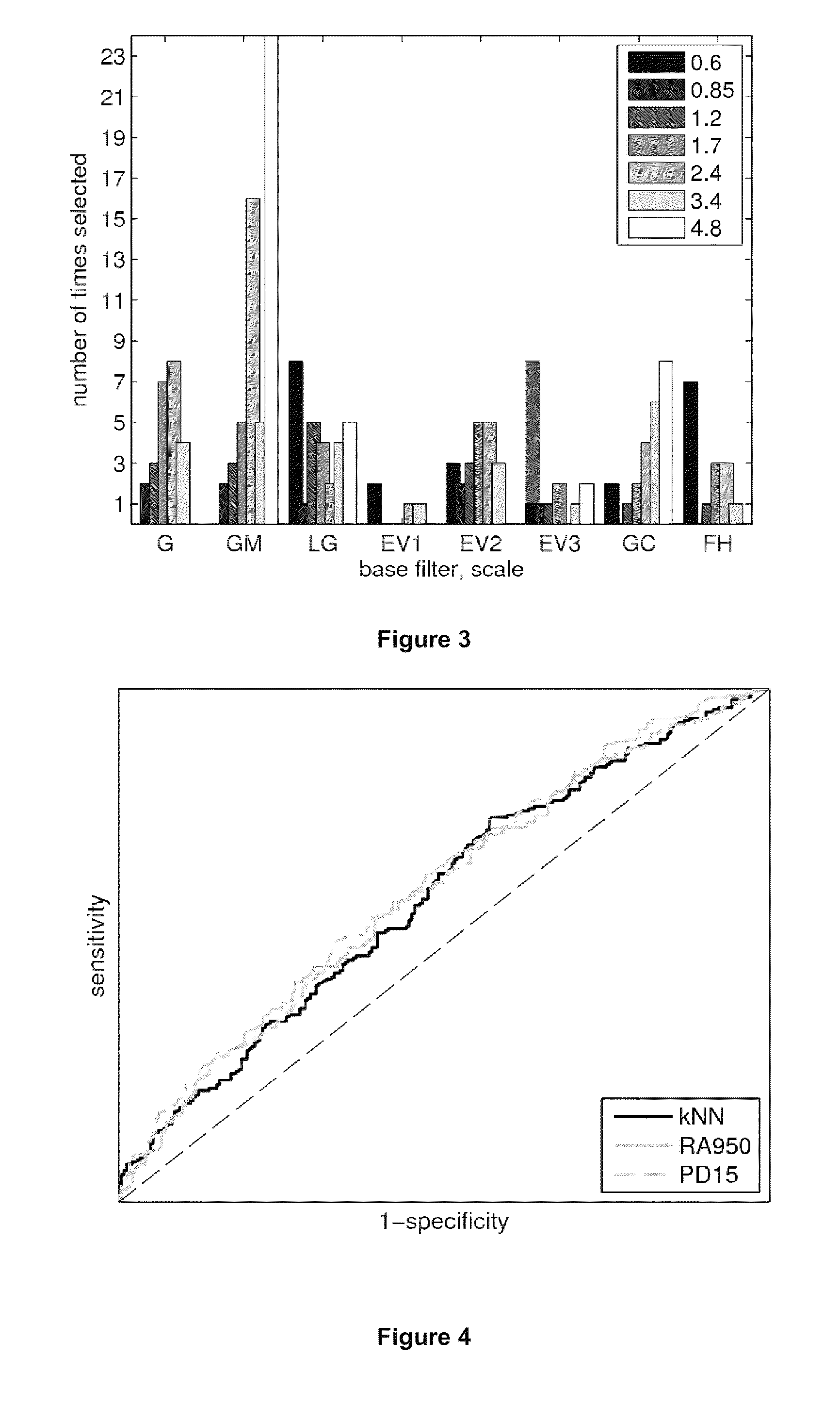
[0012]The present invention provides a method for the computerised classification of a medical diagnostic image of a lung or of a part of a lung, comprising applying to said image a trained statistical classifier which has been trained by supervised learning on a training set of methodologically similar lung images each of which images has been labelled by metadata indicative of the likelihood of the respective image relating to a lung characterised by a lung disease, or the degree of such disease, or propensity to develop such disease, wherein in said training of the classifier, for each image in the training set a number of regions of interest (ROIs) were defined, and textural information relating to the intensities of locations within each ROI was obtained, and combinations of features of said textural information were found which suitably classify said training set images according to said metadata,

[0013]and wherein, in applying said trained statistical classifier to said image,...


PUM


Abstract

The invention provides methods for automated classification of a medical diagnostic image of a lung according to its deduced probability of relating to a lung of a patient who is suffering from a diffuse parenchymal lung disease such as chronic obstructive pulmonary disease (COPD), cystic fibrosis, or severe asthma, or to a class of patients characterized by the severity of such a condition, or to a class of patients characterized by a prognostic likelihood of developing such a condition or severity of condition.

Description

CROSS REFERENCE TO RELATED APPLICATION[0001]This application claims priority to U.S. application Ser. No. 61 / 333,513, filed May 11, 2010. The entire contents of the aforementioned patent application is hereby incorporated herein by this reference.BACKGROUND OF THE INVENTION[0002]Current quantitative measures of chronic obstructive pulmonary disease (COPD) are limited in several ways. Pulmonary function tests (PFTs) are the gold standard for assessment and diagnosis of COPD [23]. These are cheap and fast to acquire but are limited by insensitivity to early stages of COPD and lack of reproducibility [8]. Visual and computerized assessment in computed tomography (CT) imaging has emerged as an alternative that directly can measure the two components of COPD, namely, small airway disease and emphysema. However, it is difficult to assess disease severity and progression visually. Moreover, visual assessment is subjective, time-consuming, and suffers from intra-observer and inter-observer ...

Claims


Application Information

Patent Timeline
Patent Type & Authority Patents(United States)
IPC IPC(8): G06K9/00G06K9/62
CPCG06T7/0012G06T7/401G06T2207/10072G06T2207/10116G06T2207/20076G06T2207/20081G06T2207/30061G06T7/44
Inventor NIELSEN, MADSDE BRUIJNE, MARLEENSORENSEN, LAUGE
Owner UNIVERSITY OF COPENHAGEN
Learn More