
Systems and methods for noninvasive analysis of retinal health and function

a retinal health and function and non-invasive technology, applied in the field of system and method for evaluating the health and function of the retina, can solve the problems of no device that can provide adequate assessment of the health and function of the photoreceptors and retinal pigment epithelium cells, and significant loss of visual ability or even blindness, and severely hinder the advancement of research, diagnosis, treatment, and patient management in connection with retinal diseases and conditions

Active Publication Date: 2015-04-21
CALIFORNIA INST OF TECH
View PDF5 Cites 4 Cited by
  • Summary
  • Abstract
  • Description
  • Claims
  • Application Information

AI Technical Summary

Problems solved by technology

Unfortunately, a number of retinal diseases and conditions can cause failure or loss of photoreceptor cell or retinal pigment epithelium functionality, leading to significant loss of visual ability or even blindness.
For some diseases, there is no device that can provide adequate assessment of the health and functionality of the photoreceptors and retinal pigment epithelium cells.
This has severely hindered the advancement of research, classification, diagnosis, treatment, and patient management in connection with retinal diseases and conditions.
In particular, for example, age-related macular degeneration is a major public health problem.
It destroys a patient's sharp and central vision in and around the macular region, significantly impacting the patient's ability to perform everyday activities (e.g., reading, driving, facial recognition, etc.) and degrading the patient's quality of life.
Age-related macular degeneration currently affects over eight million people in the United States and is the leading cause of vision loss for people over 60 years old.
Not only will patients' suffer, a significant burden may also be imposed on society, for example, due to patients' loss of independence, rising health care costs associated with age-related macular degeneration medical expenses, and injuries (e.g., falling due to poor vision).
In the wet form of age-related macular degeneration, a patient's newly grown and fragile blood vessels leak blood into the retina, prohibiting clear viewing and / or damaging the retina, and potentially causing the retina to become detached from the choroid.
While treatments have been developed for the wet form of age-related macular degeneration (e.g., laser surgery, photodynamic therapy, and anti-VEGF injunctions), there is presently no FDA-approved treatment for the dry form of age-related macular degeneration.
However, such diagnosis and classification is not adequate because the presence of drusen is ubiquitous for people over 50 years old and is considered to be associated with the natural aging process.
Accordingly, only considering the presence and amount of drusen is inadequate for researching, classifying, diagnosing or treating the disease.
Indeed, there is no adequate system or method for researching, classifying, diagnosing or treating the age-related macular degeneration.
However, because drusen is a part of the natural aging process, fundus photography images of drusen alone is not sufficient for complicated diseases such as age-related macular degeneration.
While this technique is useful in the context of wet form age-related macular degeneration, it has little applicability to other diseases such as, for example, dry form age-related macular degeneration.
However, optical coherence tomography can be sensitive to the change of the retinal structures and is not specific to the function of the retina.
Although a functional optical coherence tomography technique has been developed, its sensitivity is low and its specificity to the retina health is unclear.
Although this technique may be informative, recent histology studies show that the autofluorescence of retinal pigment epithelium cells is not necessarily correlated with the loss of photoreceptor cells.

Method used

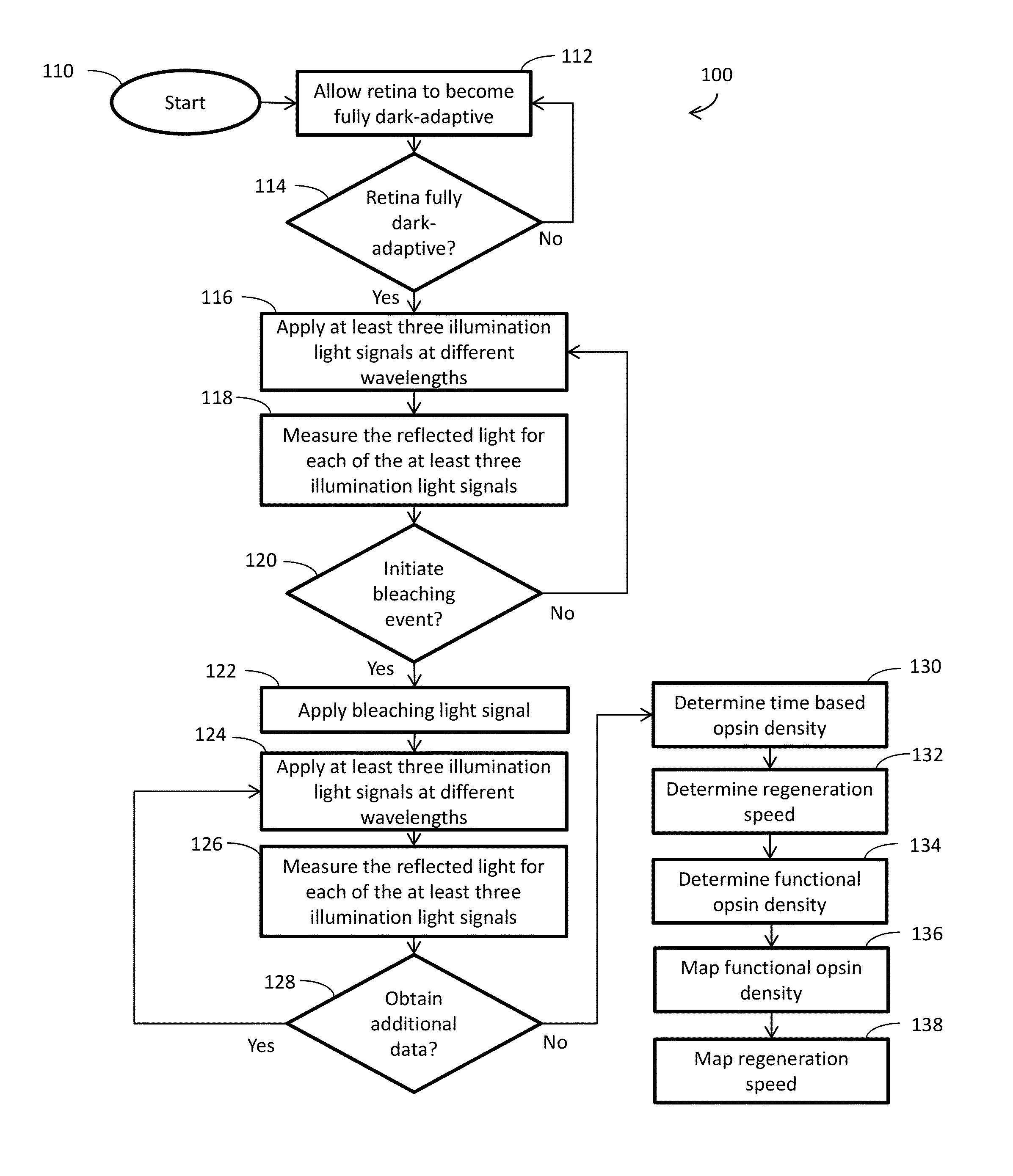

Image

  • Systems and methods for noninvasive analysis of retinal health and function
  • Systems and methods for noninvasive analysis of retinal health and function
  • Systems and methods for noninvasive analysis of retinal health and function

Examples


Embodiment Construction

[0027]While this invention is susceptible of embodiment in many different forms, there is shown in the drawings and will herein be described in detail preferred embodiments of the invention with the understanding that the present disclosure is to be considered as an exemplification of the principles of the invention and is not intended to limit the broad aspect of the invention to the embodiments illustrated. For purposes of the present detailed description, the singular includes the plural and vice versa (unless specifically disclaimed); the words “and” and “or” shall be both conjunctive and disjunctive; the word “all” means “any and all”; the word “any” means “any and all”; and the word “including” means “including without limitation.”

[0028]According to aspects of the present disclosure, systems and methods employ imaging multispectral reflectometry to directly, quantitatively, and noninvasively map the density and regeneration speed of functional photopigment (e.g., opsins such a...


PUM


Abstract

A method for noninvasive analysis of a retina includes exposing the retina to one or more first sets of at least three illumination light signals. The at least three illumination light signals each have a different wavelength. The method also includes optically collecting a reflected light signal for each of the at least three illumination light signals of the one or more first sets. Each of the reflected light signals is a portion of the respective illumination light signal reflected by the retina. The method further includes detecting the reflected light signals of the one or more first sets as a function of intensity. The method still further includes determining a first opsin density using the detected intensity of each of the reflected light signals of the one or more first sets.

Description

CROSS-REFERENCE TO RELATED APPLICATION[0001]This application claims priority of U.S. Ser. No. 61 / 562,970 filed Nov. 22, 2011, the contents of which are herein incorporated by reference.FIELD OF THE INVENTION[0002]The present invention relates generally to systems and methods for evaluating an eye and, more particularly, to systems and methods for an evaluation of health and functionality of a retina.BACKGROUND OF THE INVENTION[0003]The sense of sight involves receiving and focusing electromagnetic energy in the visible spectrum onto a retina of an eye, which converts the electromagnetic energy into signals that can be processed by the nervous system. The process by which photons of light are converted includes a cyclic decomposition and reconstitution of various proteins and enzymes within the photoreceptor cells of the retina. The photoreceptor cells include an opsin (e.g., rhodopsin in rod cells or photopsin in cone cells) bound to 11-cis retinal, a photosensitive derivative of vi...

Claims


Application Information

Patent Timeline
Patent Type & Authority Patents(United States)
IPC IPC(8): A61B3/10A61B3/00A61B3/12
CPCA61B3/12A61B3/10
Inventor CUI, XIQUANFRASER, SCOTT E.
Owner CALIFORNIA INST OF TECH
Learn More