System and method for prediction of respiratory motion from 3D thoracic images

a technology of respiratory motion and 3d thoracic image, which is applied in the field of 3d thoracic image prediction of respiratory motion, can solve the problems of difficult to accurately determine the shape, size and position of organs or lesions, and difficult to accurately estimate the 3d lung deformation, etc., to achieve the effect of improving image reconstruction

Active Publication Date: 2016-06-28
SIEMENS HEALTHCARE LTD +2
View PDF20 Cites 10 Cited by
  • Summary
  • Abstract
  • Description
  • Claims
  • Application Information

AI Technical Summary

Benefits of technology

[0007]The present invention provides a method and system for predicting respiratory motion from 3D thoracic images. Embodiments of the present invention utilize a patient-specific, generative model of the respiratory system estimated from two 3D thoracic images, such as computed tomography (CT) or magnetic resonance (MR) images. Embodiments of the present invention utilize a model that relies on a pressure force field estimated from two images at different instants in the respiratory cycle to drive the lung motion. As the model is generative, it can be used to predict the 3D respiratory motion for different respiratory patterns by modulating the pressure force field. As such, embodiments of the present invention can predict the lung position at any time of the respiratory cycle, and thus can provide a 3D surrogate for improved image reconstruction or therapy delivery.

Problems solved by technology

Medical imaging and non-invasive interventions often suffer from complications caused by respiratory motion, which is a source of artifacts in images and makes it difficult to correctly determine the shape, size, and position of organs or lesions, such as lung tumors.
Yet, because of the complexity in the respiratory motion, accurate estimation of 3D lung deformation is extremely challenging.
However, difficulties arise when the breathing pattern changes, which results in a non-periodic surrogate signal and causes imaging artifacts due to the combination of different breathing states.
Both approaches have drawbacks, such as the increase in radiation dose to achieve oversampling, or the increase of treatment or imaging time.
Furthermore, for imaging, interpolation due to the lack of information between respiratory phases can cause step artifacts.

Method used


Image

  • System and method for prediction of respiratory motion from 3D thoracic images
  • System and method for prediction of respiratory motion from 3D thoracic images
  • System and method for prediction of respiratory motion from 3D thoracic images

Examples


Embodiment Construction

[0023]The present invention relates to prediction of respiratory motion from 3D thoracic images. Embodiments of the present invention are described herein to give a visual understanding of the methods for predicting respiratory motion. A digital image is often composed of digital representations of one or more objects (or shapes). The digital representation of an object is often described herein in terms of identifying and manipulating the objects. Such manipulations are virtual manipulations accomplished in the memory or other circuitry / hardware of a computer system. Accordingly, is to be understood that embodiments of the present invention may be performed within a computer system using data stored within the computer system.

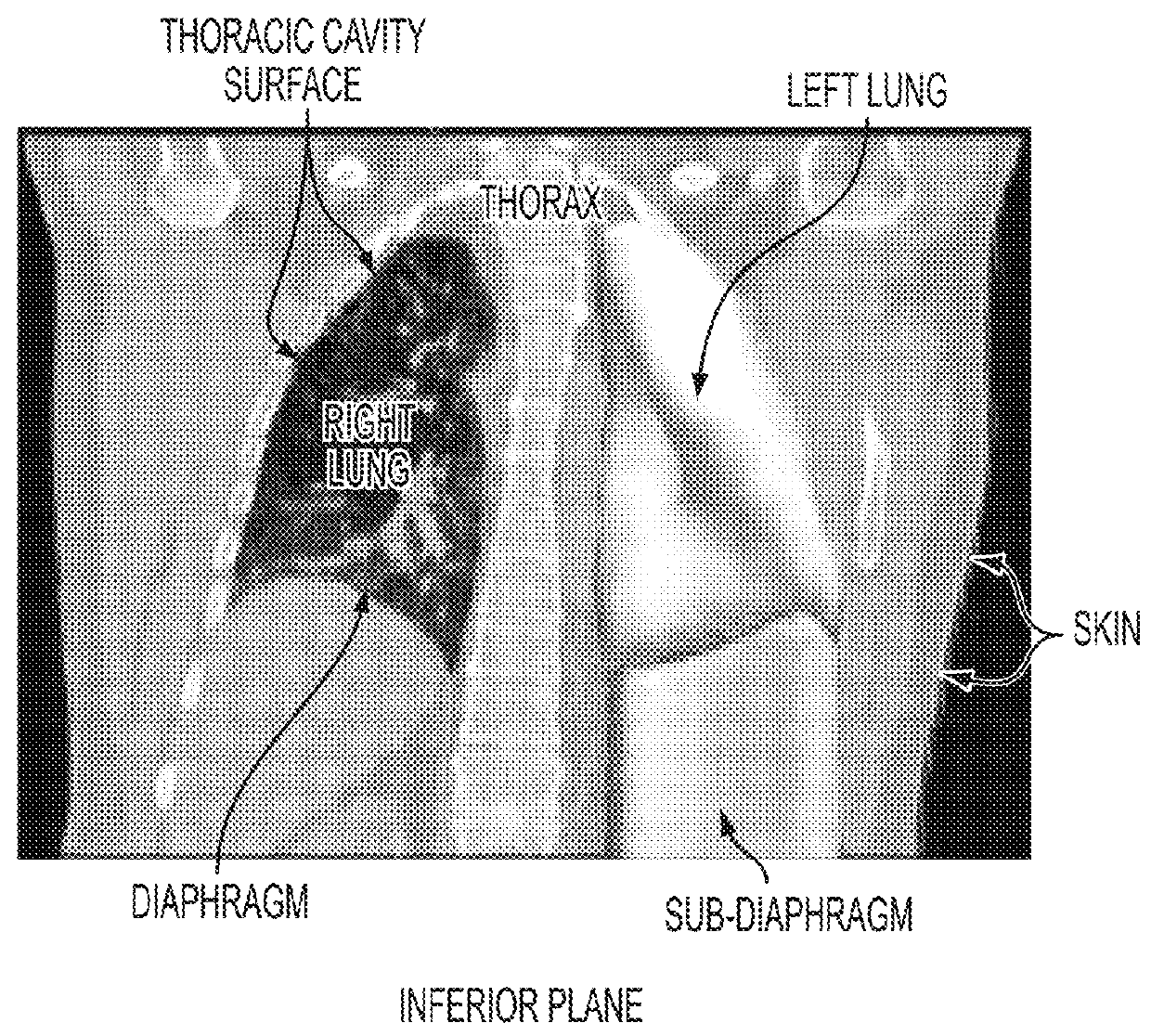
[0024]FIG. 1 is a computed tomography (CT) image showing the respiratory system of a patient. Respiratory motion is complex, as the lungs do not just compress and deform, but also slide along the thoracic cavity. The diaphragm and intercostal muscles expand th...


PUM


Abstract

A method and system for prediction of respiratory motion from 3D thoracic images is disclosed. A patient-specific anatomical model of the respiratory system is generated from 3D thoracic images of a patient. The patient-specific anatomical model of the respiratory system is deformed using a biomechanical model. The biomechanical model is personalized for the patient by estimating a patient-specific thoracic pressure force field to drive the biomechanical model. Respiratory motion of the patient is predicted using the personalized biomechanical model driven by the patient-specific thoracic pressure force field.

Description

[0001]This application claims the benefit of U.S. Provisional Application No. 61 / 876,877, filed Sep. 12, 2013, the disclosure of which is herein incorporated by reference in its entirety.[0002]The invention described was supported by National Institute of Health Award Number U01CA140206 from the National Cancer Institute.BACKGROUND OF THE INVENTION[0003]The present invention relates to prediction of respiratory motion from 3D thoracic images and more particularly to predicting deformation of the lungs and surrounding organs during respiration based on thoracic images for improved image reconstruction.[0004]Medical imaging and non-invasive interventions often suffer from complications caused by respiratory motion, which is a source of artifacts in images and makes it difficult to correctly determine the shape, size, and position of organs or lesions, such as lung tumors. The respiratory motion is complex, as the lungs do not just compress and deform, but also slide along the thoracic...

Claims


Application Information

Patent Timeline
Patent Type & Authority Patents(United States)
IPC IPC(8): G06K9/00A61B5/113G06T7/20G06F19/00A61B5/00A61B5/11G16Z99/00
CPCA61B5/7275A61B5/113A61B5/1128G06F19/3437G06T7/2046G06T2207/10072G06T2207/30061G06T7/251G16H50/50G16Z99/00
Inventor BOETTGER, THOMASCARNIS, FRANCOISCOMANICIU, DORINDECLERCK, JEROMEFUERST, BERNHARDKAMEN, ALIMANSI, TOMMASOZHANG, JINGDAN
Owner SIEMENS HEALTHCARE LTD
Why Eureka
Try Eureka