Detection of infectious disease in a human or animal by measuring specific phagocytosis in a thin film sample of their anticoagulated blood

a technology of anticoagulation blood and target analyte, which is applied in the direction of instruments, biochemical equipment and processes, material analysis, etc., can solve the problem of inconclusive test results, and achieve the effect of optimal phagocytosis and easy detection

Inactive Publication Date: 2016-07-19
ABBOTT POINT CARE
View PDF9 Cites 26 Cited by
  • Summary
  • Abstract
  • Description
  • Claims
  • Application Information

AI Technical Summary

Benefits of technology

[0007]The method of this invention utilizes an electronic photometric microscopic analysis of the blood sample involving electronic imaging or scanning of the sample. The method involves placing the incubated blood sample, as described below, in a test chamber of predetermined and fixed thickness so as to produce a thin layer of the blood sample in the chamber. At least one wall of the chamber is transparent so that the sample can be observed in the chamber. In certain cases both the top and bottom walls of the chamber are transparent. The thickness of the chamber can range from 3μ to 25μ. When anticoagulated whole blood is being analyzed the chamber should be preferably 4μ-6μ thick so as to obtain a thin film monolayer of the blood sample, which monolayer contains both white blood cells and red blood cells. The monolayer blood sample in the chamber will be imaged for the presence or absence of white blood cells that have bound or ingested the first detectable particles which are coated with an antigen that is specific to the target Lyme disease analyte, i.e., a Borrelia bacteria. The blood sample will also contain a similar number of second detectable particles, identical in size and composition to the first particles except for distinguishable color or fluorescence, which are not coated with a Lyme disease analyte-specific antigen. Lyme disease is used only as an example of one disease that may be detected using this method and when the term “Lyme” is used any infectious disease process that is characterized by alterations in phagocytosis may be substituted. The first and second particles will be characterized by different detectable signals such as color and or fluorescence so that they can be distinguished one from the other. The particles should otherwise be the same in size and composition, differing only by the presence or absence of the target antigen or antigens, since size and composition can affect phagocytosis. The particles to be used as type one and type two particles are both ideally identically sized in a range from about 1 to 3 microns in diameter, which is a size that permits easy detection and optimal phagocytosis by white blood.

Problems solved by technology

If no particles or too few particles (defined as a number below the user defined normal range) of both type one particles and type two particles are phagocytosed, the test is invalid and such invalidity may be due dead or incompetent white blood cells or inadequate incubation conditions.
If greater amounts of type one particles than type two particles are ingested but both amounts are within the normal range the test is inconclusive.
Such a result can have important clinical utility but may not be used to indicate the presence or absence of the specific disease process being tested for.

Method used


Image

  • Detection of infectious disease in a human or animal by measuring specific phagocytosis in a thin film sample of their anticoagulated blood
  • Detection of infectious disease in a human or animal by measuring specific phagocytosis in a thin film sample of their anticoagulated blood

Examples


Embodiment Construction

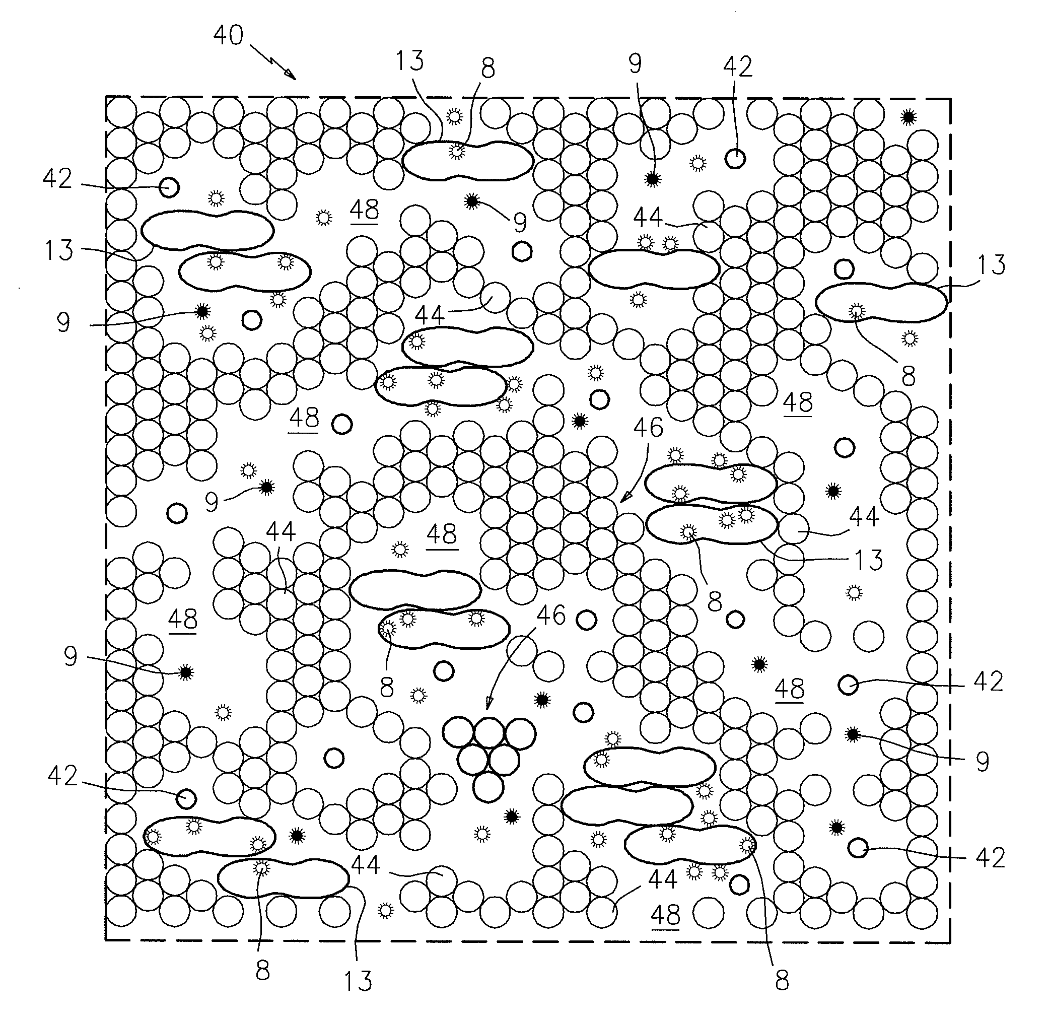
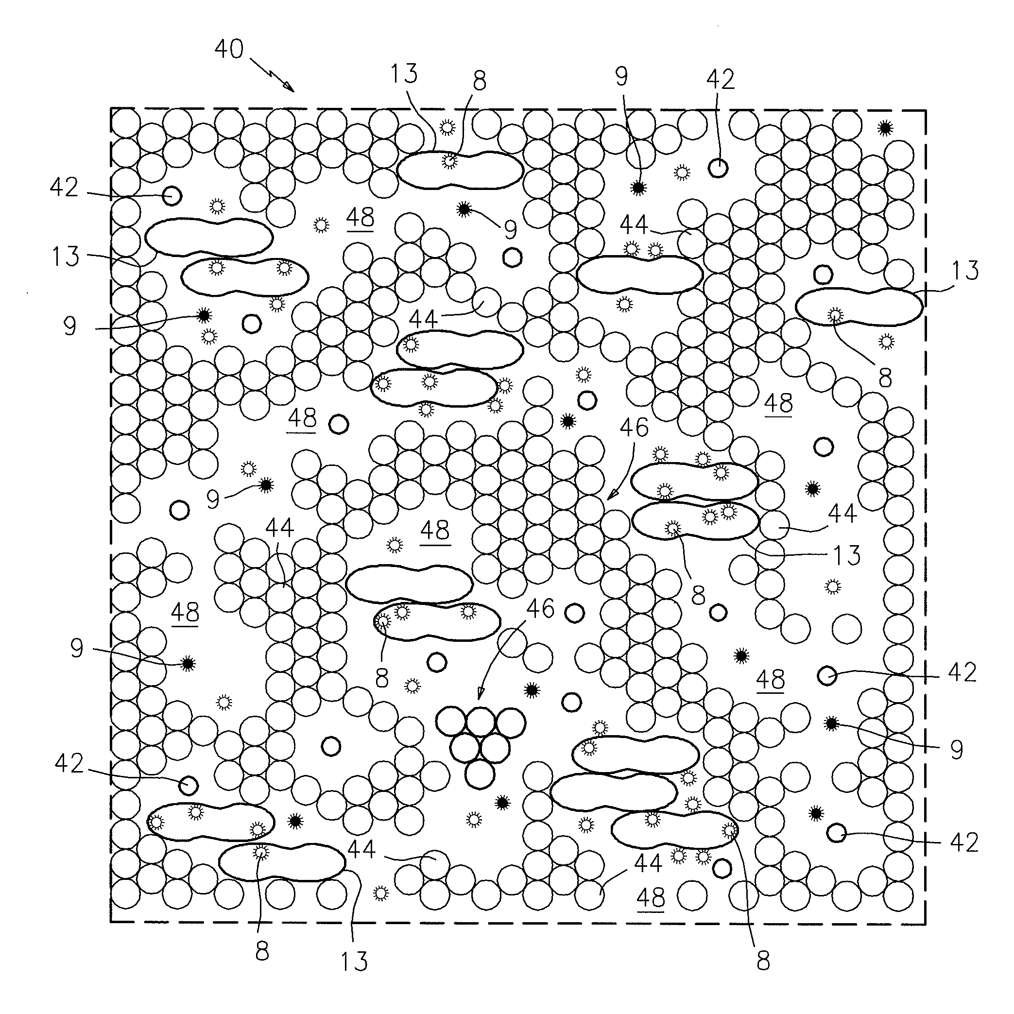
[0011]Referring now to FIG. 1, there is shown in plan view a portion of a sampling chamber 40 which contains a thin monolayer film sample of anticoagulated whole blood that has been incubated with the detection particles. In this case, the chamber 40 has a transparent upper surface so that the blood sample can be electronically or photometrically imaged. The thin film blood sample is contained in a sampling chamber which is provided with beads 42 that provide the desired thickness of the chamber 40, which in this case is about 6μ. The thin film of blood includes red blood cells 44, red blood cell clumps or rouleaux 46, plasma cell-free lacunae 48, and white blood cells 13. The blood sample also includes two detectable particles 8 and 9. The particles 8 are coated with the antigen or antigens specific to the target Lyme disease analyte, and the particles 9 are not coated with the target analyte-specific antigen(s). The particles 8 and 9 are differentially detectable in the sample whe...


PUM

PropertyMeasurementUnit
through plane thicknessaaaaaaaaaa

Abstract

A method for performing a target analyte assay of a thin film anticoagulated blood sample, wherein the target analyte is the presence or absence of specific phagocytosis and / or binding of particles coated with a particular antigen or antigens by white blood cells present in the anticoagulated blood sample, wherein the particles are coated with the particular antigen or antigens, which antigens are similar or identical to antigens expressed by a defined pathogenic infectious agent of interest.

Description

BACKGROUND OF THE INVENTION[0001]1. Technical Field[0002]This invention relates to a method for performing a target analyte assay of a thin film anticoagulated blood sample, where the target analyte is the presence or absence of specific phagocytosis and / or binding of particles coated with a particular antigen or antigens by white blood cells, which blood sample contains the white blood cells present in the anticoagulated blood sample, and detectable first type particles coated with the particular antigen or antigens present on the surface which antigens are similar or identical to the antigens expressed by the defined pathological agent. More particularly, the present invention relates to electronic detection, quantization and characterization of phagocytosis and / or binding of detectable substances in human and animal biological fluids such as whole blood, or other fluids containing white blood cells, which characterization is performed in a thin chamber on a quiescent thin film fl...

Claims


Application Information

Patent Timeline
Patent Type & Authority Patents(United States)
IPC IPC(8): C12Q1/00C12Q1/68G01N33/543G01N33/53G01N30/00G01N33/569C12Q1/04G01N33/50G01N15/14G01N15/00
CPCG01N33/56911C12Q1/04G01N33/5094G01N33/5432G01N15/1475G01N2015/008Y02A50/30
Inventor LEVINE, ROBERT A.WARDLAW, STEPHEN C.
Owner ABBOTT POINT CARE
Why Eureka
Try Eureka