Visualization of anatomical data by augmented reality

a technology of anatomical data and augmented reality, applied in the field of system and method for visualizing anatomical data, can solve the problems of prone to failure, limited application range, and limited scope of medical imaging modalities, and achieve the effect of enhancing user flexibility and avoiding lengthy initialization

Active Publication Date: 2016-11-22
DEUTES KREBSFORSCHUNGSZENT STIFTUNG DES OFFENTLICHEN RECHTS
View PDF7 Cites 41 Cited by
  • Summary
  • Abstract
  • Description
  • Claims
  • Application Information

AI Technical Summary

Benefits of technology

[0008]It is hence the objective of the present invention to provide an improved system and method for visualizing anatomical data that avoids a lengthy initialization and provides enhanced user flexibility.

Problems solved by technology

However, only few medical imaging modalities are capable of providing real-time images of the patient's anatomy.
However, in such conventional techniques it is usually the task of the physician to mentally transfer the three-dimensional virtual image to the patient, or in other words to establish a correspondence between the real object and the 3D virtual image generated from the static medical data.
This not only requires considerable skill and experience, but is also prone to failures, which might have very serious consequences for the patient under therapy.
In addition, navigation in the three-dimensional data set may not be as intuitive as desired and is often rather cumbersome.
However, the system disclosed in US2007 / 0018975 A1 requires means adapted to track the pose of the camera in real space, and hence can only be employed in specially equipped environments, e.g. in a specially equipped operating theatre.
In addition, the patient is not allowed to move during inspection, for otherwise the coincidence of the coordinate system of the virtual model with the coordinate system of the camera in real space may be lost.

Method used


Image

  • Visualization of anatomical data by augmented reality
  • Visualization of anatomical data by augmented reality
  • Visualization of anatomical data by augmented reality

Examples


Embodiment Construction

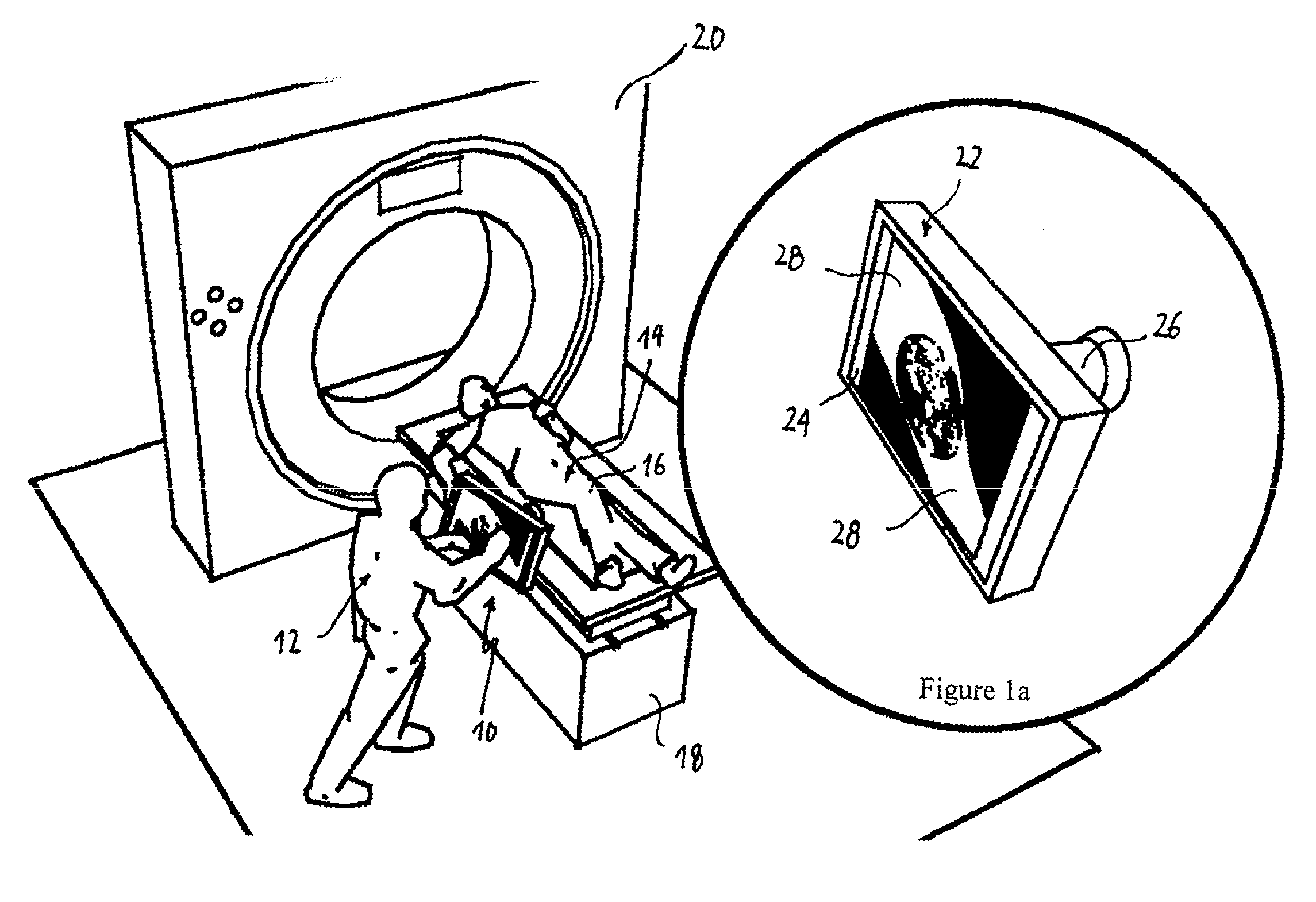
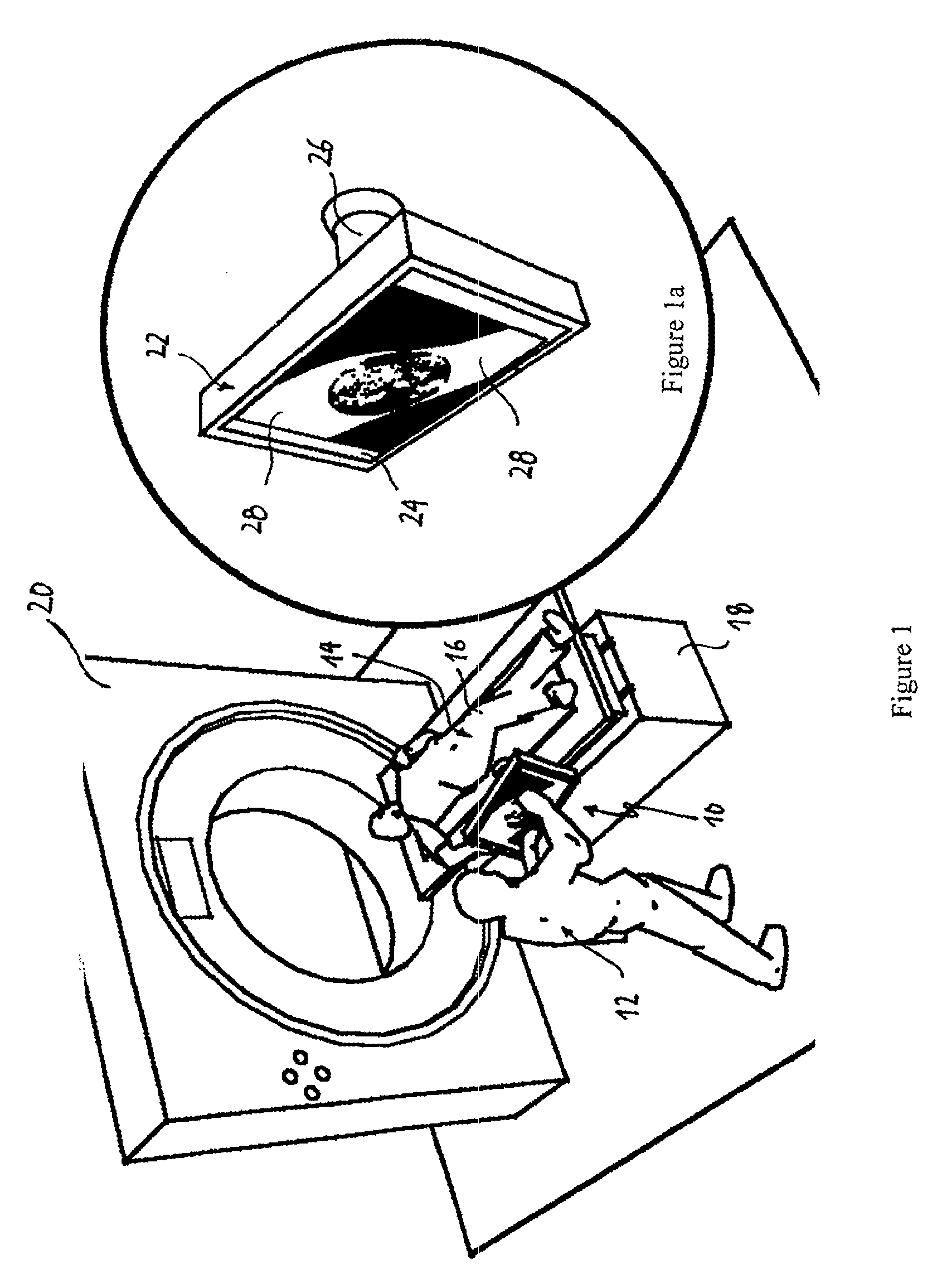
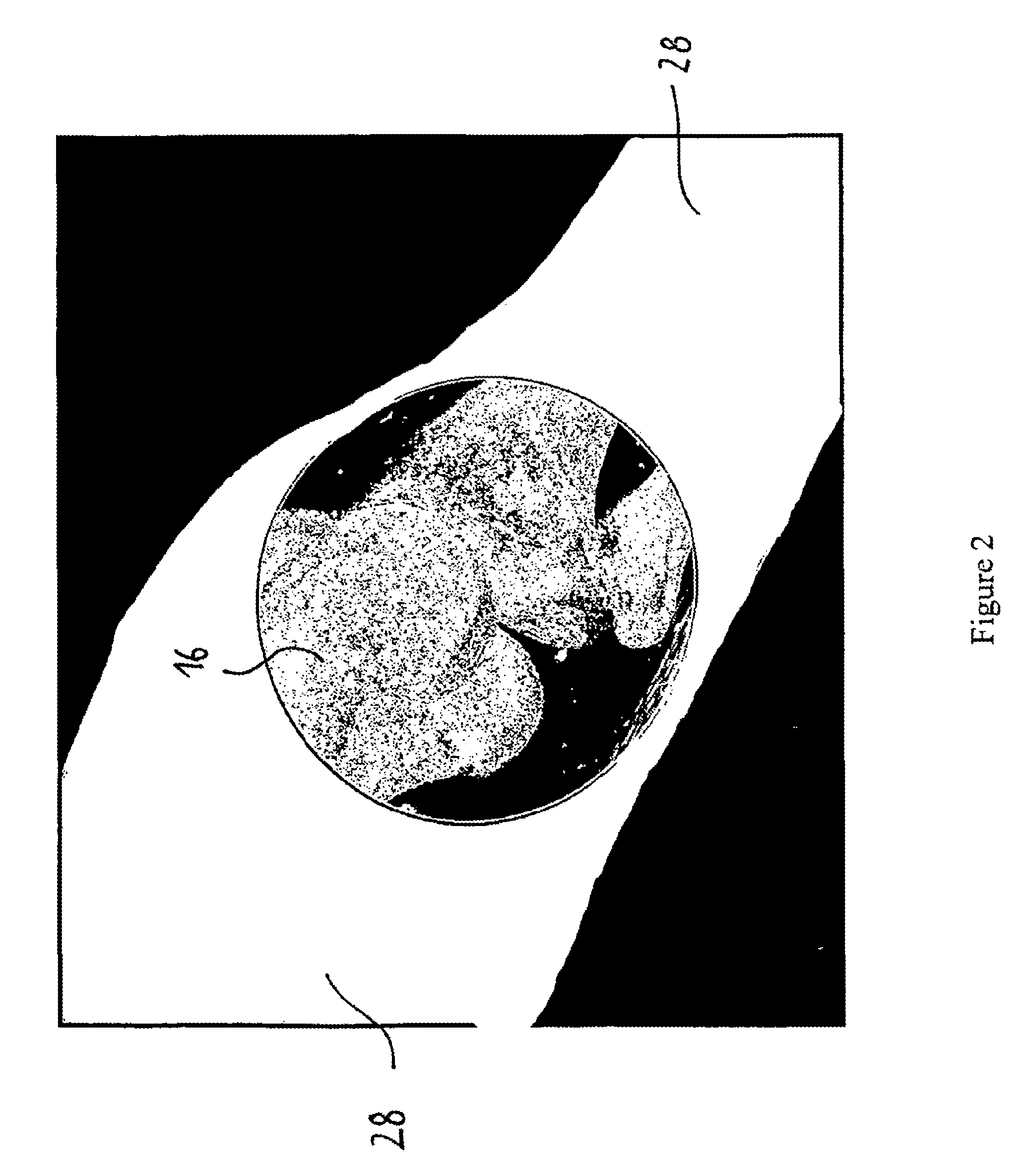
[0100]FIG. 1 illustrates a visualization system 10 according to the present invention as used by a physician 12 to examine the knee joint 16 of a patient 14. The patient 14 is drawn resting on a movable table 18 in an examination room equipped with a computed tomography scanner 20. However, this is a mere example, and it is one of the advantages of the present invention that the visualization system 10 can be employed in almost any environment, be it in a hospital or at the patient's home.

[0101]The visualization system 10 according to the specific example is illustrated in greater detail in FIG. 1a and comprises a display means 22 equipped with a screen 24. The display means 22 shown in FIG. 1 is a tablet PC, but may alternatively be any other computer device equipped with a screen 24 and suitable for displaying images. Mounted and physically attached to the backside of the display means 22 (opposite to the screen 24) is a time-of-flight camera 26. The time-of-flight camera 26 may a...


PUM


Abstract

A sensor means is employed to sense a distance to the surface of a subject to be examined, so that a range image may be acquired. Intensity information may be acquired alongside the distance information. The distance information and intensity information may be evaluated to track the pose of the sensor means relative to the surface of the subject to be examined, so that anatomical data related to said subject may be displayed as seen from the position and / or orientation of the sensor means or display means. By moving the sensor means or display means along the surface of the subject to be examined, such as a patient in a hospital environment, the user hence gets the impression of being able to look directly into the human body.

Description

[0001]This application is a continuation of co-pending Patent Cooperation Treaty PCT / EP2011 / 005735, filed Nov. 14, 2011, which in turn claims priority from European Patent Application 10191085.9, filed Nov. 12, 2010, which are incorporated herein by reference.TECHNICAL FIELD[0002]The present invention relates to a system and method for visualizing anatomical data, in particular to a system and method for mobile augmented reality for on-patient visualization of pre-recorded medical images and to a system and method for assisting in image-guided interventions.BACKGROUND ART[0003]Visualization of anatomical data for disease diagnosis, surgical planning, or orientation during interventional therapy is an integral part of modern health care. However, only few medical imaging modalities are capable of providing real-time images of the patient's anatomy. A common procedure therefore involves the acquisition of static three-dimensional images, e.g. by means of computed tomography (CT) or ma...

Claims


Application Information

Patent Timeline
Patent Type & Authority Patents(United States)
IPC IPC(8): A61B5/00A61B6/00
CPCA61B5/0035A61B5/0077A61B5/742A61B34/10A61B34/20A61B90/361A61B6/462A61B6/5223A61B6/5235A61B2034/105A61B2034/2065A61B2090/061A61B2090/365A61B2090/372
Inventor MAIER-HEIN, LENAFANGERAU, MARKUSMEINZER, HANS-PETERSEITEL, ALEXANDER
Owner DEUTES KREBSFORSCHUNGSZENT STIFTUNG DES OFFENTLICHEN RECHTS
Why Eureka
Try Eureka