Convolutional neural networks for cancer diagnosis

Active Publication Date: 2017-08-22
ANIXA DIAGNOSTICS CORP
View PDF31 Cites 58 Cited by
  • Summary
  • Abstract
  • Description
  • Claims
  • Application Information

AI Technical Summary

Benefits of technology

[0016]In some embodiments, detecting the presence or absence of the target cell in the method of diagnosing cancer comprises a computer-implemented method comprising: (a) transmitting, by a computer, a surface scanning instruction to a multichannel fluorescence acquisition device, wherein the microfilter is positioned on a stage of the multichannel fluorescence acquisition device; the surface scanning instruction comprising one or more commands for the multichannel fluorescence acquisition device to determine the z-axis height of the microfilter surface; (b) transmitting, by a computer, an image capture instruction to the multichannel fluorescence acquisition device, the image capture instruction comprising one or more commands for the multichannel fluorescence acquisition device to acquire a plurality of digital images of the microfilter using a channel corresponding to the fluorophore, wherein the plurality of digital images are acquired at each stage position in a z-sta

Problems solved by technology

High numbers of TAMs, in the stroma of a tumor, are

Method used


Image

  • Convolutional neural networks for cancer diagnosis
  • Convolutional neural networks for cancer diagnosis
  • Convolutional neural networks for cancer diagnosis

Examples


example 1

tration of a Biological Sample

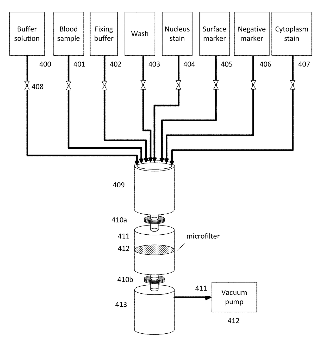
[0284]Blood samples from cancer and normal patients were drawn for enrichment of tumor derived cells using microfiltration. The microfilter comprised nickel-palladium and had the following structural features: 13 mm diameter, 8 um pore size, and 8 um thickness. The microfilter was assembled into a cassette comprising a fitting for an input syringe and a retention syringe.

[0285]For each patient, a 7.5 mL sample of the patient's blood was mixed with fixation buffer. The cassette was attached to a retention syringe, which was subsequently inserted into a syringe pump. An input syringe was then attached to the cassette. All washes and materials applied to the membrane within the cassette were applied through the input syringe. The microfilter was washed with phosphate buffered saline (PBS). The fixed patient blood sample was transferred to the input syringe and the blood sample was drawn from the input syringe, through the microfilter, and into the retentio...

example 2

of Microfiltration-Isolated Cells from a First Panel of Breast Cancer Patients

[0286]A first panel of 4 breast cancer patients had their blood drawn, filtered and stained as described in Example 1. Microfilters corresponding to blood samples from each patient were scanned at 40× in order to determine whether TDMCs were observed using the microfilters.

[0287]Patient Breast-101 (right lumpectomy, right SNB) was diagnosed with breast cancer. Blood was drawn prior to surgery for a right lumpectomy and right sentinel node biopsy. After blood sample filtering, staining, and microscopic filter membrane imaging at 20× and 40× in a four channel fluorescent microscope, 6 image sets (each image set includes four individual fluorescence channels and a composite) from approximately 500 frame sets were selected for detailed observation. At least 3-4 TDMCs were identified in the images.

[0288]Observation of filtered cells from patient Breast-101 demonstrated cells that were DAPI positive (blue channe...

example 3

of Microfiltration-Isolated Cells from Normal Patients

[0296]A panel of normal patients had their blood drawn, filtered and stained as described in Example 1. Microfilters corresponding to blood samples from each patient were scanned at 40× in order to determine whether TDMCs were observed using the microfilters.

[0297]Normal Subject Female Normal-001F: Blood was Drawn from Normal Subject N-001F. After blood sample filtering, staining, and microscopic filter membrane imaging at 20× and 40× in a four channel fluorescent microscope, approximately 500 frame sets were analyzed. As shown by the sample image of FIG. 21, no TDMCs were found in this sample.

[0298]Normal Subject Female Normal-002F: Blood was drawn from normal subject N-002F. After blood sample filtering, staining, and microscopic filter membrane imaging at 20× and 40× in a four channel fluorescent microscope, approximately 500 frame sets were analyzed. As shown by the sample image of FIG. 22, no TDMCs were found in this sample....


PUM


Abstract

The present disclosure provides devices and systems for diagnosing and characterizing cancer in a subject. Devices include microfilters and microfiltration systems useful for the isolation and characterization of cells from a subject.

Description

CROSS-REFERENCE[0001]This application claims the benefit of U.S. provisional application Ser. No. 62 / 308,763, filed Mar. 15, 2016, which is incorporated by reference herein in its entirety.BACKGROUND OF THE INVENTION[0002]In the course of a lifetime, cancer will strike about one in three women and one in two men. More than 560,000 die from it each year in the United States alone. Early detection and treatment is currently the leading method to reduce cancer death, especially if the cancer is detected before metastasis. For nearly all types of cancer, the 5-year relative survival rate is substantially lower if the disease is caught at an advanced stage. Moreover, the financial costs of cancer treatment at an advanced stage can be an additional burden. By 2020, the cost of cancer treatment is expected to be $207 billion annually in the United States. Accordingly, early detection of cancer is important for increasing cancer survival rates and reducing the cost of treatment.[0003]Howeve...

Claims


Application Information

Patent Timeline
IPC IPC(8): G01N33/574G01N21/64
CPCG01N33/57492G01N33/57415G01N33/57449G01N21/6428G01N21/6458G01N2333/70589G01N2333/4742G01N2333/705G01N2021/6439G01N2201/12G01N33/574G01N33/6893
Inventor KUMAR, AMITROOP, JOHN
Owner ANIXA DIAGNOSTICS CORP
Why Eureka
Try Eureka