Corneal epithelial sheet and process for producing the same

A production method and technology of epithelial sheets, applied in the fields of biochemical equipment and methods, epidermal cells/skin cells, nervous system cells, etc.

Inactive Publication Date: 2007-08-08
阿如布勒斯特有限公司 +1
View PDF2 Cites 10 Cited by
  • Summary
  • Abstract
  • Description
  • Claims
  • Application Information

AI Technical Summary

Problems solved by technology

However, in monocular diseases (corneal erosion, etc.), it has been reported that the corneal epithelium of the healthy eye is transplanted into the affected eye to obtain good results. However, most of the refractory corneal diseases are binocular, so in fact can't use this method

Method used


Image

  • Corneal epithelial sheet and process for producing the same
  • Corneal epithelial sheet and process for producing the same
  • Corneal epithelial sheet and process for producing the same

Examples


Embodiment 1

[0106]

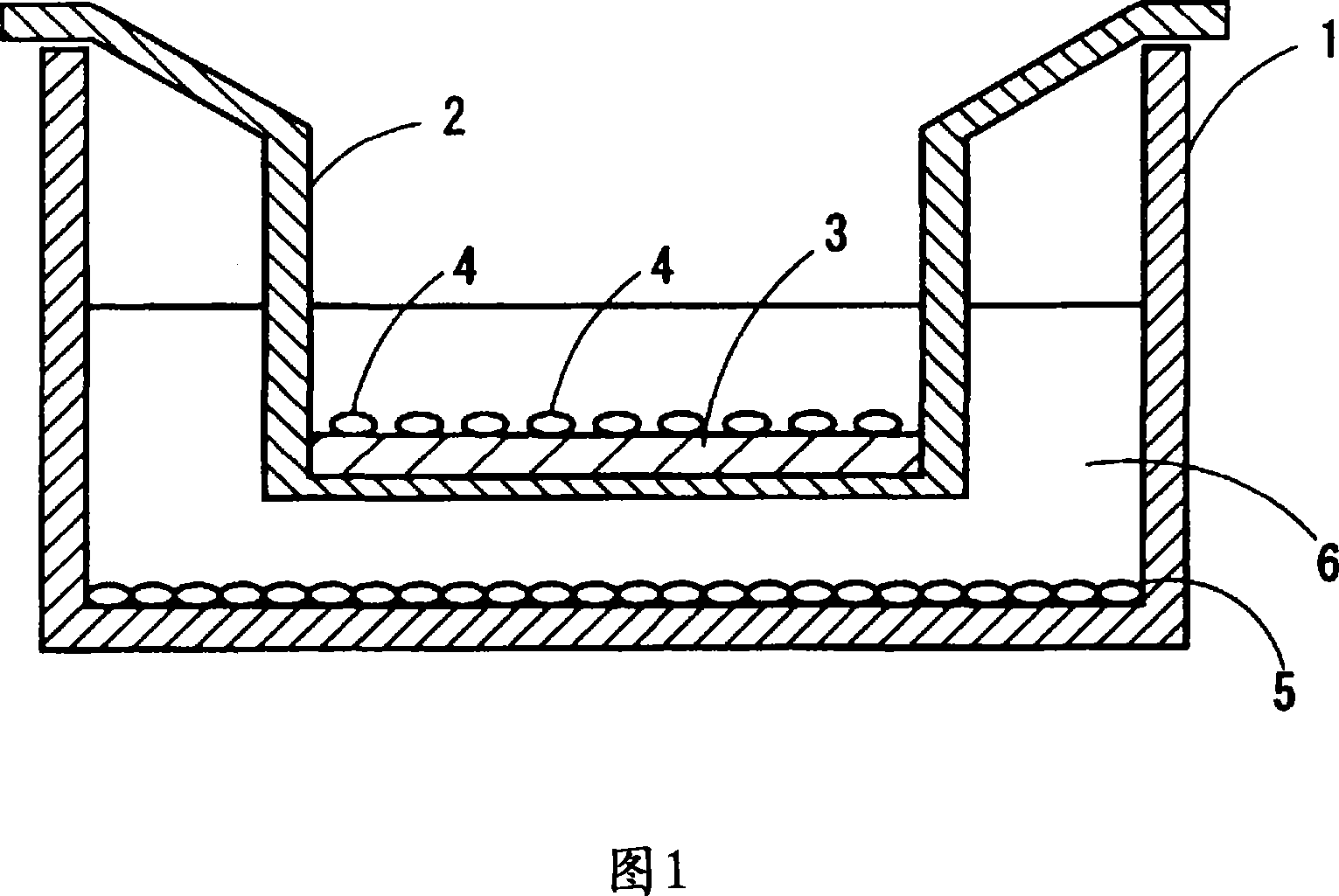
[0107] 1-1 Collection of amniotic membrane

[0108] For pregnant women who do not have systemic complications and are about to undergo caesarean section, amnion membranes will be taken during caesarean section in the operating room after a careful explanation with the obstetrician and gynecologist in advance, and after obtaining consent. The operation should pay attention to cleanliness, wash hands according to the operation operation, and then put on the surgical gown. Preparing for delivery Clean the amnion taken with an enamel dish and wash with saline. After delivery the placental tissue is moved to an enamel tray and the amniotic tissue is separated from the placenta by hand. The firmly healed portion of the amnion and placenta are excised with scissors.

[0109] 1-2. Treatment of amniotic membrane

[0110] The process of amnion treatment is carried out in the order of (1) cleaning, (2) trimming, and (3) preservation. During the whole process, it is prefera...

Embodiment 2

[0151]

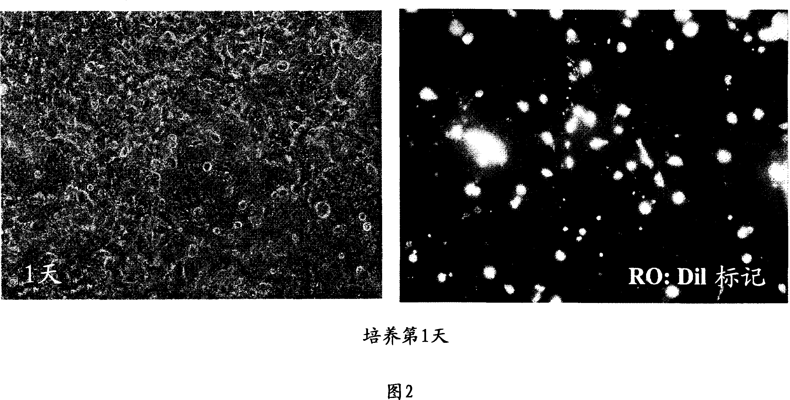
[0152] Human oral mucosal epithelial cell suspensions were prepared from healthy volunteers after obtaining consent in the same manner as in the method described in 1-5. (Recovery of oral mucosal epithelial cells) above. Separately, corneas obtained from the Northwest eye bank were treated in the same manner as described in 1-6. (Recovery of corneal epithelial cells) above to obtain a suspension of human corneal epithelial cells. The human oral mucosal epithelial cells and human corneal epithelial cells prepared in this way were co-cultured with 3T3 cells using the human amniotic membrane with scratched epithelium as the matrix. A corneal epithelial-like cell layer is formed. The preparation method of scratched human amniotic membrane and the co-culture conditions with 3T3 were as described above in 1-1. (Collection of amniotic membrane) to 1-4. (Treatment of amniotic membrane epithelium).

[0153] The results of the morphology, organization, and immunostaining of ...

Embodiment 3

[0156]

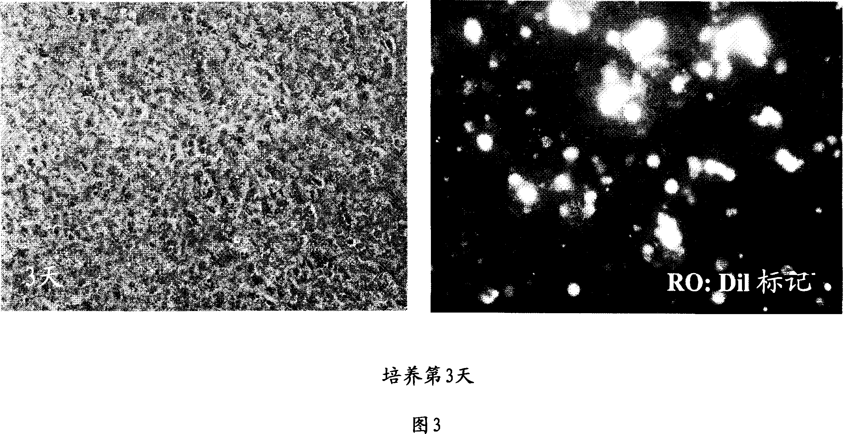
[0157]Oral mucosal epithelial cell suspensions were prepared from healthy volunteers after obtaining consent in the same manner as described in 1-5. (Recovery of oral mucosal epithelial cells) above. In addition, human amnion was obtained by the method described in 1-1. (Collection of amnion) and 1-2. (Processing of amnion) above. Thereafter, the epithelial cells (suspension of human amniotic epithelial cells) were separated by treating with 0.05% trypsin-EDTA solution (GIBCOBRL) for 15 minutes. The human oral mucosal epithelial cells and human amniotic membrane epithelial cells prepared in this way were co-cultured with 3T3 cells using the amniotic membrane with scratched epithelium as the matrix, and after about 14 days of culture (including 3 days of culture by air lifting method), the formation of Corneal epithelioid cell layer. The preparation method of scratched amniotic membrane and the conditions of co-culture with 3T3 cells were as described above in 1-1. ...


PUM


Abstract

A corneal epithelial sheet having a cell layer similar to the anterior epithelium of cornea. A corneal epithelial sheet is obtained through the step (a) of separately preparing a first cell being a self cell and a second cell whose origin is different from that of the first cell, the step (b) of planting the first cell and the second cell on a collagen layer and culturing them, and the step (c) of after proliferation of the first cell and second cell resulting in formation of a cell layer, bringing the outermost stratum of the cell layer into contact with air.

Description

technical field [0001] The invention relates to a corneal epithelial sheet and a manufacturing method thereof. The present invention can be used in the treatment of diseases requiring transplantation of corneal epithelium (ocular surface diseases). The present invention provides an effective treatment means especially for binocular corneal diseases. Background technique [0002] Corneal epithelial transplantation is currently performed as a surgical treatment for ocular surface diseases in which the cornea is covered with conjunctival epithelium and cloudiness occurs. However, the prognosis for refractory keratoconjunctival diseases with severe inflammation (Stevens-Johnson syndrome or ocular pemphigoid, corneal erosions, etc.) is very poor. The biggest reason is that the allo corneal epithelium with strong antigenicity is recognized by the host's immune system and rejected. In particular, complication caused by systemically or locally administering a large amount of immu...

Claims


Application Information

Patent Timeline
Patent Type & Authority Applications(China)
IPC IPC(8): A61L27/00A61F2/14
CPCC12N2501/01C12N5/0621C12N2501/33C12N2501/11A61L27/3891C12N2502/097C12N2533/92A61L27/3813A61F2/14A61L27/00
Inventor 木下茂中村隆宏
Owner 阿如布勒斯特有限公司
Why Eureka
Try Eureka