

Quick testing strip for residual pesticide
A technology for testing tylosin and test strips, which is applied in the direction of measuring devices, instruments, scientific instruments, etc., can solve the problems of false detection and missed detection, achieve low investment, wide application range, and are not easy to false negative and false positive misjudgment Effect
- Summary
- Abstract
- Description
- Claims
- Application Information
AI Technical Summary
Problems solved by technology
Method used
Image

Examples
preparation example Construction
[0029] 4. Preparation of goat anti- or rabbit anti-mouse IgG
[0030]Extract mouse serum IgG with saturated ammonium sulfate, take 1 part of mouse serum and 2 parts of PBS (pH 7.2) and mix well, add an equal volume of saturated ammonium sulfate and mix well, put in 4°C refrigerator for 2h, centrifuge at 4°C, 1200r / min for 15min , discard the supernatant; dissolve the precipitate with an appropriate amount of PBS (pH7.2), add saturated ammonium sulfate to a final concentration of 33%, place in a refrigerator at 4°C for 2h, centrifuge at 1200r / min at 4°C for 15min, discard the supernatant, and dissolve the precipitate with a small amount of PBS (pH7.2). .2) Dissolve the precipitate, put it in a refrigerator at 4°C and dialyze with PBS (pH7.2) overnight, change the medium 2 to 3 times, centrifuge at 12,000 r / min at 4°C for 15 minutes, collect the supernatant, and measure the protein concentration with a UV spectrophotometer , immunize healthy sheep or rabbits with 50-100 μg / kg bo...
Embodiment 1
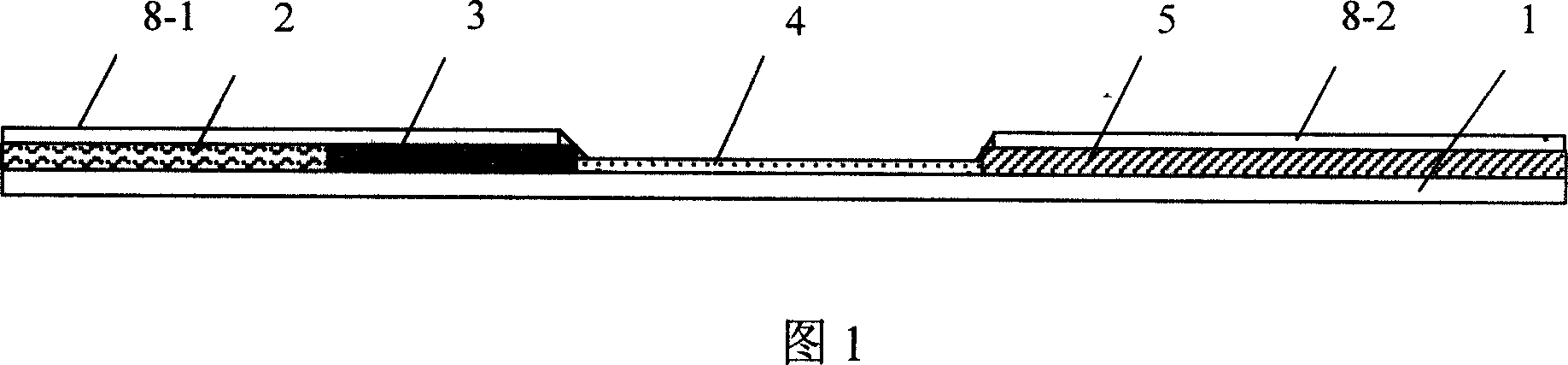
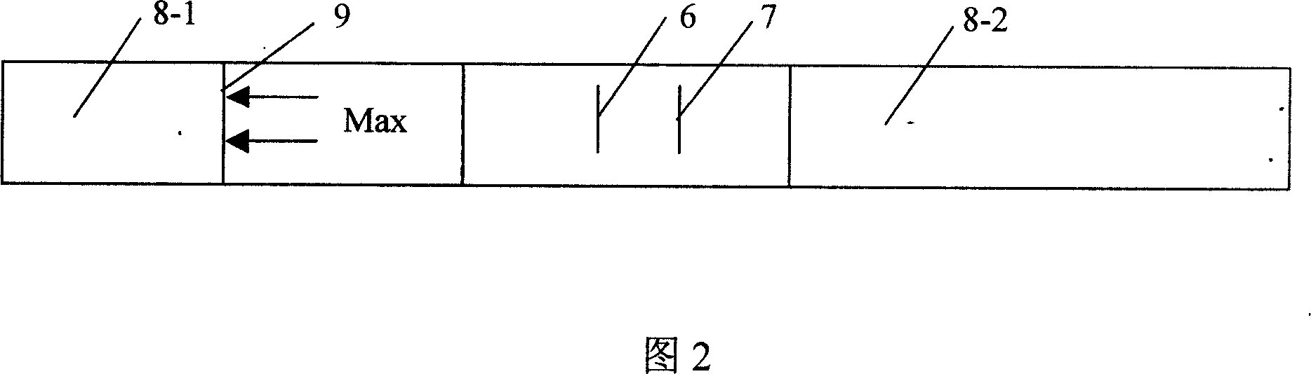
[0033] Example 1: The structure of the test strip for rapid detection of tylosin residues is shown in Fig. 1 and Fig. 2 . Among the figure 1 is the support layer, made of plastic strips, 2 is the fiber layer at the test sample end, made of glass wool, 3 is the gold-labeled antibody (W1) fiber layer adsorbed with tylosin TL, according to the above specific implementation The preparation method described in mode 3 is to prepare gold-labeled antibody (W1) glass wool adsorbed with tylosin monoclonal antibody. Made of filter paper, the layers numbered 2, 3, 4, and 5 are pasted and fixed on the plastic strip 1 in sequence from left to right, and the fibers at the junction of each layer interpenetrate with each other. On the nitrocellulose membrane layer 4, 6 is the detection blot "|" printed with tylosin-conjugated bovine serum albumin (BSA) solution, and 7 is the control printed with goat anti-mouse IgG (Y) solution Imprint "|", two imprints are arranged in parallel to form a comb...
Embodiment 2
[0038] Embodiment 2: The structure of the test strip for rapid detection of tylosin residues is basically the same as that of Embodiment 1, the difference is that: the support layer 1 is made of non-absorbent hard paper strips, and the fiber layer 2 of the test end is made of nylon membrane, gold Colloidal gold-labeled tylosin TL polyclonal antibody is adsorbed on the labeled antibody fiber layer 3, pure cellulose membrane is used in the cellulose film layer 4, ferritin is the carrier protein solution coupled to TL in the invisible detection blot zone 6, and the invisible control blot Band 7 was prepared on cellulose membrane with rabbit anti-mouse IgG solution.
[0039] Preparation of testing samples: for testing meat samples, the samples are chopped and ground, and the ratio of meat samples and saline is 1:30 to make the sample suspension to be tested.
[0040] Other operating methods and result judgments are the same as those in Example 1 and will not be repeated.
PUM
| Property | Measurement | Unit |
|---|---|---|
| diameter | aaaaa | aaaaa |
Abstract
Description
Claims
Application Information