Digital medical image analysis

A medical image and image technology, applied in the field of digital medical image analysis, can solve problems such as increasing the complexity of CAD software, achieve high sensitivity or specificity, and reduce time constraints

Inactive Publication Date: 2007-12-05
MEDICSIGHT
View PDF5 Cites 9 Cited by
  • Summary
  • Abstract
  • Description
  • Claims
  • Application Information

AI Technical Summary

Problems solved by technology

Allowing users to set parameter values ​​increases the complexity of CAD software

Method used


Image

  • Digital medical image analysis
  • Digital medical image analysis
  • Digital medical image analysis

Examples


Embodiment Construction

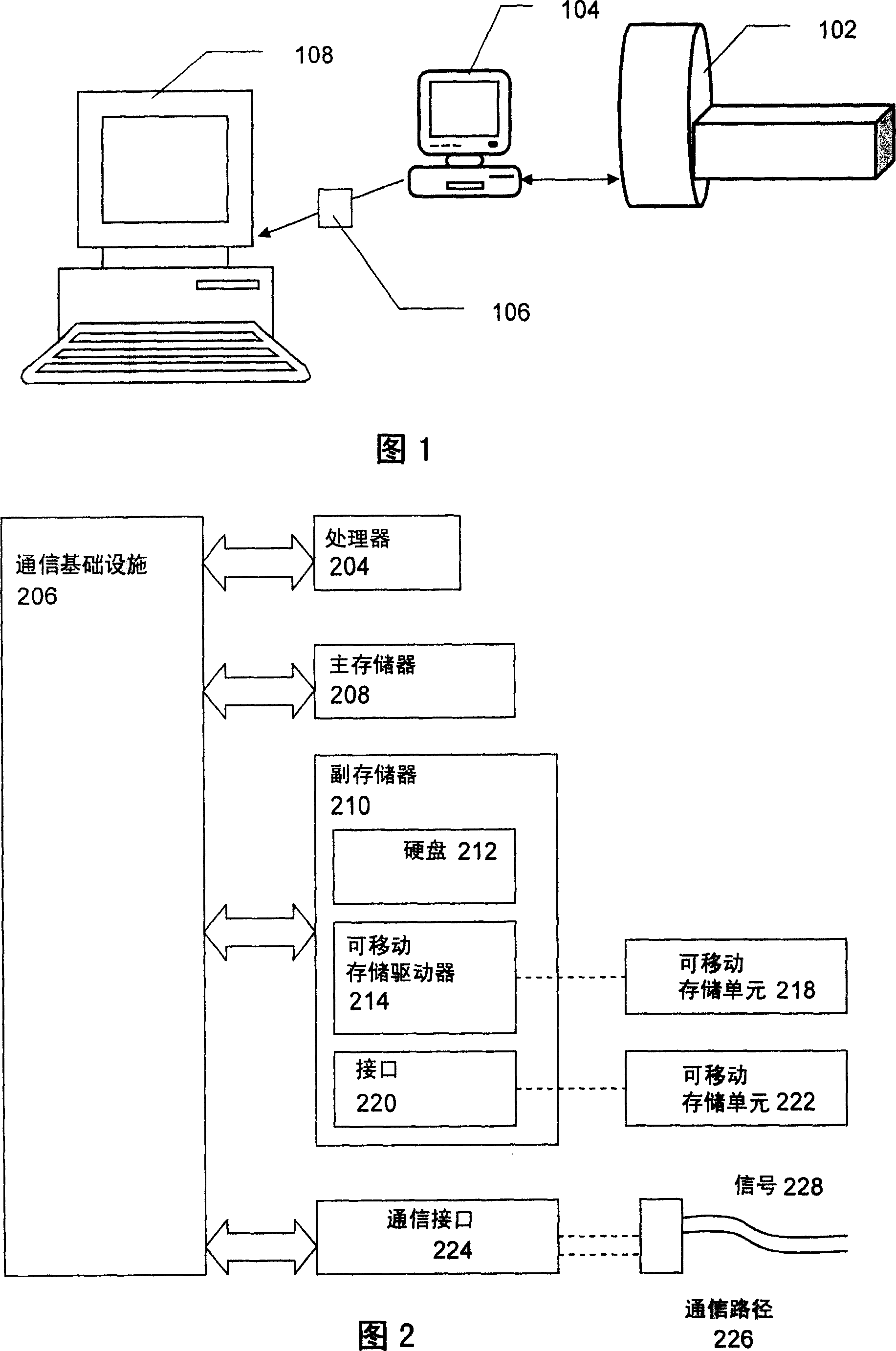
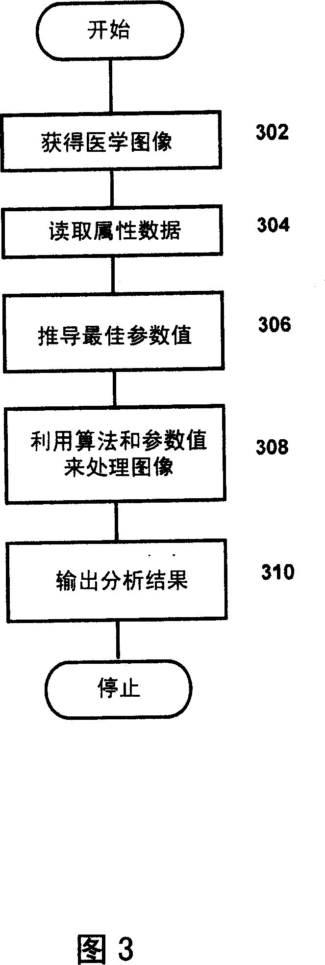
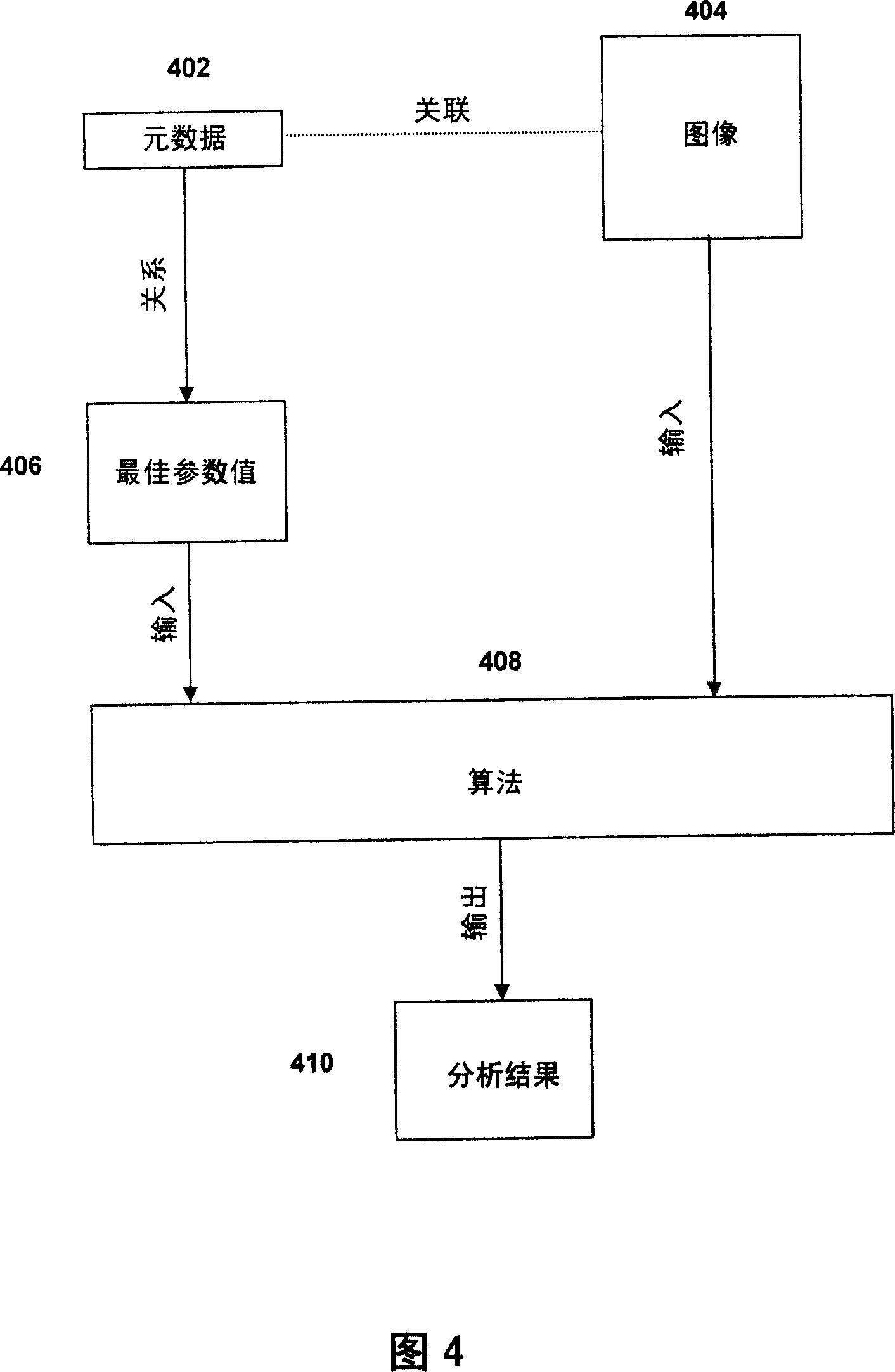
[0038] Medical Image Acquisition

[0039] The invention is applicable to digital medical images. An example of such an image is a CT scan image. A CT scan image is a digital image comprising one or a series of CT image slices obtained from a CT scan of an area of ​​a patient or diseased animal. Each slice is a 2D digital grayscale image of the X-ray absorption of the scanned area. The properties of the slices depend on the CT scanner used; for example, a high-resolution multi-slice CT scanner can produce images with a resolution of 0.5-0.6 mm / pixel in the x and y directions (ie, in the slice plane). Each pixel has 32-bit grayscale resolution. The intensity value of each pixel is normally expressed in HU. Serial slices can be separated by a constant distance along the z-direction (scan separation axis); for example, by a distance of 0.75-2.5 mm. Thus, the scanned image may be a three-dimensional (3D) grayscale image, the total size of which depends on the area and number o...


PUM


Abstract

A medical image (404) is analyzed using an algorithm (408) requiring input parameters (406), the values of which are derived from metadata (402) indicating properties of the image (404). The metadata may indicate the type of image acquisition device (102) or the settings used to create the image (404), and/or relate to the patient from whom the image (404) was taken. This allows optimum values to be selected for the parameters.

Description

technical field [0001] The present invention relates to digital medical image analysis, especially digital medical image analysis by means of computer-implemented algorithms. Background technique [0002] Medical images are valuable tools for detection, diagnosis, and evaluation, as well as surgical planning, for disease treatment. A variety of physical techniques and modalities have been developed to produce these images, including projection X-rays, computed tomography (CT), ultrasound (US), positron emission tomography (PET), and magnetic Resonance Imaging (MRI). Traditionally, these images are reviewed by trained radiologists or other clinicians. These images can be generated digitally (US, CT and MRI), or can be digitized from analog images (film). Radiologists or clinicians can review digitized images, annotate them, and generate reports. All result data is stored for later retrieval and analysis. [0003] The user's task is made easier by examining images with an...

Claims


Application Information

Patent Timeline
Patent Type & Authority Applications(China)
IPC IPC(8): G06F19/00G06T5/00G16H30/40
CPCA61B6/583G06F19/321G06F19/345G06T7/604G06T2207/30004G06T2207/30061G06T2200/24G06T5/00G06T7/0012G06T7/64G16H50/20G16H30/40G06T2207/20081G06T2207/20084
Inventor 贾姆希德·德默思凯苏珊·艾里森·伍德保罗·理查德·贝洛克·萨姆尔
Owner MEDICSIGHT
Why Eureka
Try Eureka