Digital blood vessel contrast image enhancement method integrating context information

An angiography and image enhancement technology, applied in the field of medical imaging, can solve the problems of limited enhancement effect, no consideration of the interaction of blood vessel space structure, and difficulty in achieving visual effects

Inactive Publication Date: 2008-08-06
HUAZHONG UNIV OF SCI & TECH
View PDF0 Cites 10 Cited by
  • Summary
  • Abstract
  • Description
  • Claims
  • Application Information

AI Technical Summary

Problems solved by technology

However, these methods mostly start from the gray level information of the local area, without considering the spatial structure of blood vessels and the interaction between contexts, so the enhancement

Method used


Image

  • Digital blood vessel contrast image enhancement method integrating context information
  • Digital blood vessel contrast image enhancement method integrating context information
  • Digital blood vessel contrast image enhancement method integrating context information

Examples


example

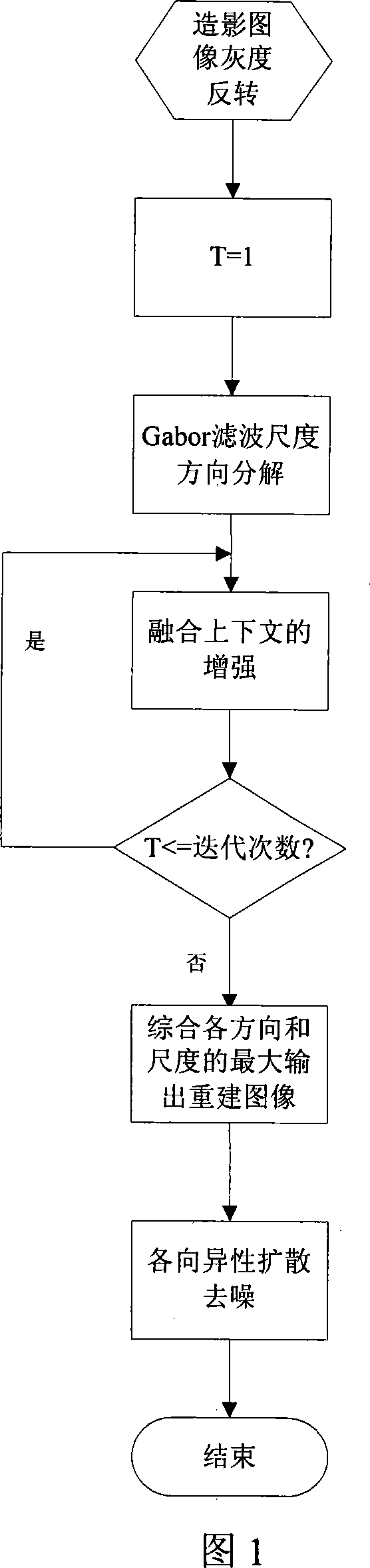
[0030] As shown in Figure 1, the process of this example is:

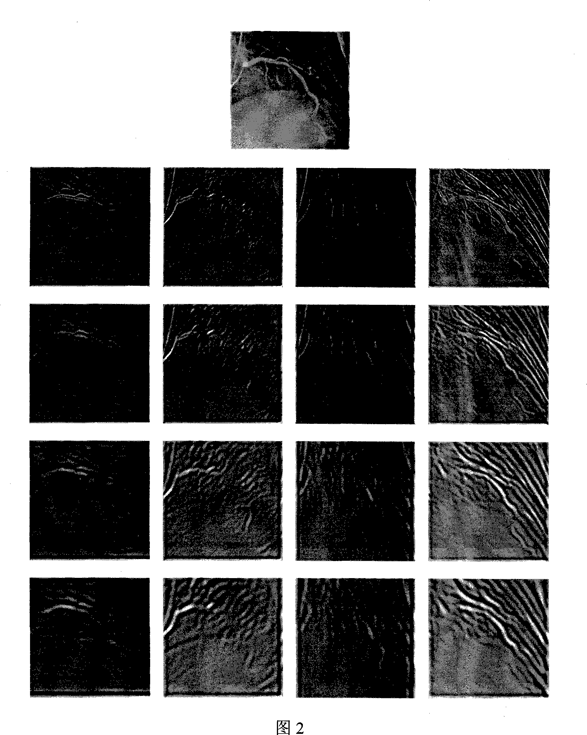
[0031] (1) Use the Hubble (Gabor) filter to decompose the grayscale inverted digital angiography image into K directions and L scales. The value of K usually ranges from 4 to 16. The number of scales L can be determined according to the largest blood vessel. Width W max and minimum vessel width W min to make sure.

[0032] (1.1) In this example, the value of K is 12, and the value of L is 4. The corresponding angle

[0033] θ = π × ( k - 1 ) K , k = 1,2 , · · · , K

[0034] Each angle represents a possible direction of enhancement.

[0035] (1.2) Different scales can be obtained by adjusting the center frequency of t...


PUM


Abstract

The invention discloses a digital angiography image enhancing method combined with context information. The invention decomposes an image to different frequencies and azimuths through a Gabor filter at first, then utilizes the structural consistency of local spaces of bloods to ensure that a consistent space ingredient is enhanced based on information that a cocircular rule is combined with blood context through an iteration process, and finally obtains a satisfactory visual effect through smooth denoising of anisotropic diffusion. A cardiovascular image is usually full of noise, and the prior enhancing technology is difficult to enhance blood vessels, in particular thin blood vessels, from the cardiovascular image with low contrast and complex background. The digital angiography image enhancing method combined with context information enhances the ingredient we are interested in according to the global feature of the target, enhances the ingredient, the blood vessel, with better space structure from the angiography image, has good local interference resistant performance, and particularly shows good enhancing performance to blood vessel endings with higher level and lower contrast.

Description

technical field [0001] The invention belongs to the technical field of medical imaging, and in particular relates to a digital angiography image enhancement method which fuses context information. Background technique [0002] As a basic image processing technology, image enhancement aims to process images to obtain images with better visual effects and more useful images for specific applications. Due to the different purposes and requirements of specific applications, the specific enhancement techniques required may also be quite different. Fundamentally speaking, there is no universal standard for image enhancement, and the observer is the ultimate judge of the quality of an enhancement technique. [0003] Angiography is a technique with a long history to visualize blood vessels. Digital Subtraction Angiography (DSA) is a widely used vascular visualization technology developed on this basis. It has been used in clinical practice for more than 20 years and is an importan...

Claims


Application Information

Patent Timeline
IPC IPC(8): G06T5/00
Inventor 唐奇伶桑农张天序王岳环李艳西
Owner HUAZHONG UNIV OF SCI & TECH
Why Eureka
Try Eureka