Three-dimensional visualization method and device

A three-dimensional and image technology, applied in the field of three-dimensional visualization, can solve problems such as difficult judgment, difficulty in medical diagnosis, disorganized internal geometric models, etc., to achieve the effect of accurately displaying spatial relationships and improving the quality of image visualization.

Inactive Publication Date: 2008-09-24
SOUTH CHINA NORMAL UNIVERSITY +1
View PDF0 Cites 41 Cited by
  • Summary
  • Abstract
  • Description
  • Claims
  • Application Information

AI Technical Summary

Problems solved by technology

Then, for the finally obtained segmented image, the small cavities may be the "imprint" of the small tissues (such as blood vessels) inside it, or it may be the "imprint" of noise, resulting in the three-dimensional analysis of each tissue after segmentation. After reconstruction, when it is necessary to transparently display the images of each layer, it is difficult to judge whether the cavity is real tissue or noise, which makes it difficult to judge the spatial position relationship between various tissues. When the noise is serious, it may even make the geometric model The interior of the car is disorganized, which makes medical diagnosis difficult

Method used


Image

  • Three-dimensional visualization method and device
  • Three-dimensional visualization method and device
  • Three-dimensional visualization method and device

Examples


Embodiment 1

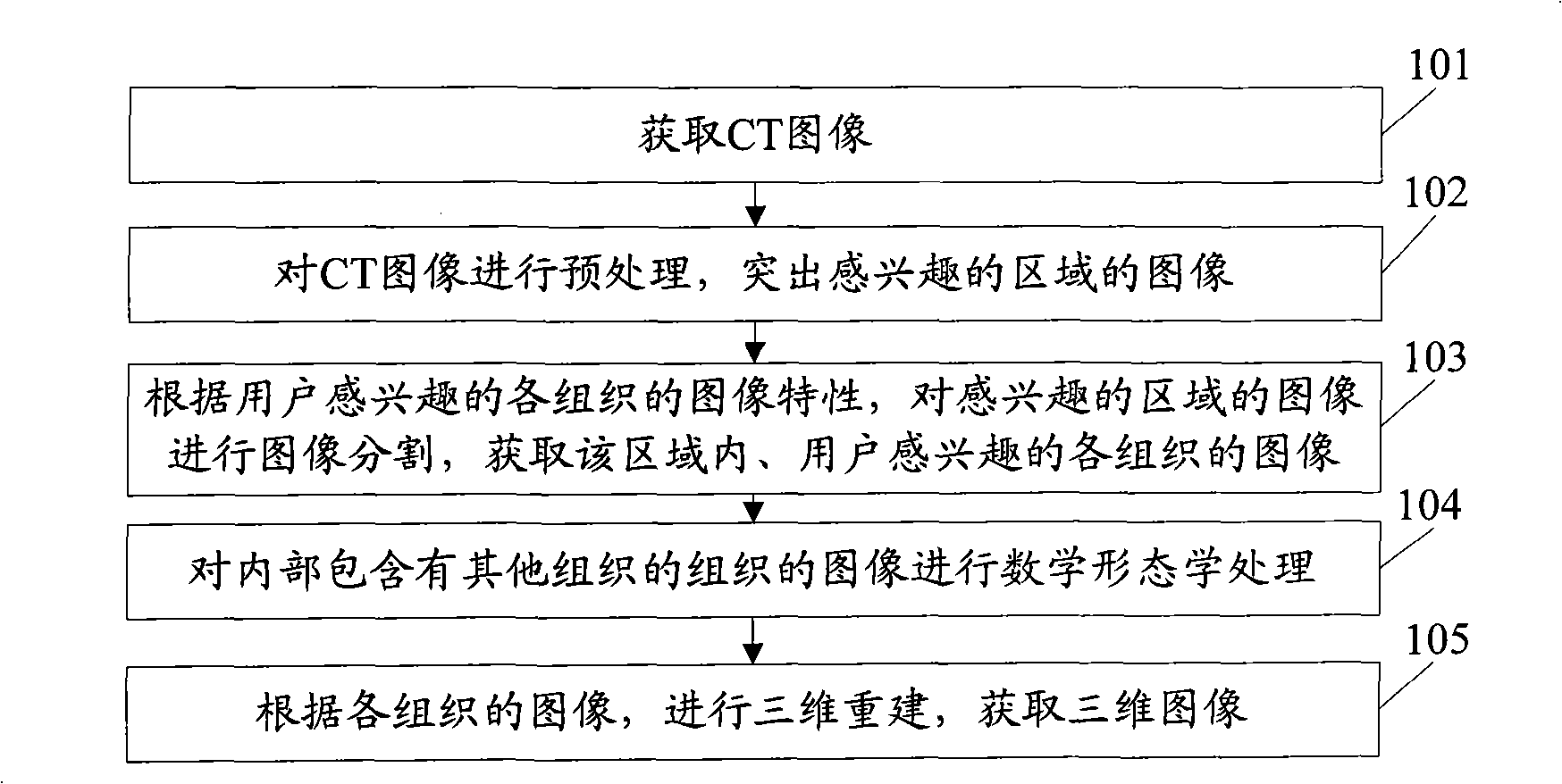
[0042] figure 1 The flow diagram of the three-dimensional visualization method provided in this embodiment, as shown in the figure, the method includes:

[0043] Step 101: Obtain a CT image.

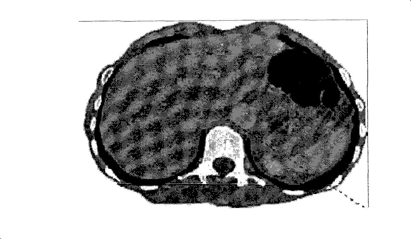
[0044] At present, medical CT data is acquired through CT. CT images are expressed in different gray levels, reflecting the degree of tissue absorption of X-rays. Therefore, like the black-and-white images shown in X-ray images, black shadows indicate low-absorption areas, that is, low-density areas, such as lungs with a lot of gas; white shadows indicate high-absorption areas, that is, high-density areas, such as bones. However, compared with X-ray images, CT has a higher density resolution, that is, a higher density resolution (density resolutiln). Therefore, although the density difference of human soft tissue is small, although the absorption coefficient is much close to that of water, it can also be imaged by contrast. Therefore, CT scan can better display the organs composed of...

Embodiment 2

[0126] Figure 12 A schematic structural diagram of a three-dimensional visualization device provided for this embodiment, as shown in the figure, the device includes:

[0127] The input unit 121 is used for inputting CT images.

[0128] For its principle, refer to the description of step 101 in Embodiment 1.

[0129] The preprocessing unit 122 is configured to preprocess the CT image input by the input unit 121 to highlight the image of the region of interest. Various existing preprocessing methods can be selected to preprocess the image to highlight the image of the region of interest.

[0130] For its principle, refer to the description of step 102 in Embodiment 1.

[0131] The segmentation unit 123 is configured to perform image segmentation on the image of the region of interest processed by the preprocessing unit 122, and acquire images of tissues of interest to the user in the region. Various segmentation methods in the prior art may be adopted as the segmentation m...

Embodiment 3

[0141] Figure 13 A schematic structural diagram of a three-dimensional visualization device provided for this embodiment, as shown in the figure, the device and Figure 12 The difference of the shown device is that the device of this embodiment may also include:

[0142] The median filtering unit 131 is configured to perform median filtering on images of tissues that do not contain other tissues inside, and input the processed images to the three-dimensional reconstruction unit 135 .

[0143] The three-dimensional reconstruction unit 135 performs three-dimensional reconstruction according to the tissue filtered by the median filter unit 131 and the tissue processed by the morphology processing unit 124 .


PUM


Abstract

The invention relates to a computer image processing field and the embodiment of the invention discloses a three-dimensional visualization method and an apparatus. The method includes: the CT image is preprocessed to highlight the image of an interested area; according to an image feature of an interested tissue, the image of the interested area is segmented to get the image of the interested tissue in the area; the tissue image comprising other tissues to be segmented inside is processed mathematically and morphologically; according to the image of every tissue, a three-dimensional reconstruction is made to obtain a three-dimensional image. The application of the technical proposal of the embodiment of the invention is capable of making the image data of a human body of an individual visualized, and helpful to show relations of spatial positions of each tissue accurately.

Description

[0001] technology neighborhood [0002] The invention relates to the field of computer image processing, in particular to a three-dimensional visualization method and device. Background technique [0003] Medical image visualization is to display human body information obtained by digital imaging technologies such as computerized tomography (CT) and magnetic resonance imaging (MRI) in a three-dimensional effect on the computer, thereby providing Structural information not available by traditional means. Because it can provide doctors with realistic three-dimensional images, it is convenient for doctors to observe and analyze from multiple angles and levels based on the provided images. Therefore, medical image visualization plays an important role in auxiliary diagnosis and medical teaching. [0004] The image visualization processing in the prior art mainly includes the following steps: [0005] Carry out segmentation processing on human body image data to obtain image data...

Claims


Application Information

Patent Timeline
IPC IPC(8): G06T5/00G06T17/00A61B6/03
Inventor 鲍苏苏方驰华彭丰平鲁朝敏徐清振
Owner SOUTH CHINA NORMAL UNIVERSITY
Why Eureka
Try Eureka