Method for acquiring complete anatomy image with segmenting pencil-beam CT image by split joint

A CT image and cone-beam technology, applied in image enhancement, image data processing, instruments, etc., can solve the problem of not being able to fully display the tumor location and shape, the inability to fully display the anatomical structure of tumor tissue, and the inability to completely reproduce the anatomical structure of the lung and other issues, to achieve the effect of improving the effect of radiation therapy, accurate and accurate dose correction, and accurate plan evaluation

Active Publication Date: 2010-07-28
AFFILIATED CANCER HOSPITAL OF SHANDONG FIRST MEDICAL UNIV SHANDONG CANCER INST (SHANDONG CANCER HOSPITAL)
View PDF3 Cites 0 Cited by
  • Summary
  • Abstract
  • Description
  • Claims
  • Application Information

AI Technical Summary

Problems solved by technology

[0008] The disadvantage of the existing technology in actual use is that the OBI equipment can only reconstruct image segments less than 14cm in length from the obtained CBCT images, and use these image segments to formulate and evaluate major cancers such as nasopharyngeal cancer, esophageal cancer, rectal cancer, and cervical cancer. For some tumor radiotherapy plans, it is not enough to fully display the position and shape of the tumor in the human body, that is, the anatomical structure of the tumor tissue cannot be fully displayed, which affects the formulation of radiotherapy plans on CBCT images; on the other hand, for chest tumors, Even if the complete tumor tissue is photographed, the anatomical structure of the entire lung is usually not fully reproduced, which will cause doctors to be unable to accurately assess the actual radiation dose of the lung tissue that is sensitive to radiotherapy when receiving radiotherapy through CBCT images

Method used


Image

  • Method for acquiring complete anatomy image with segmenting pencil-beam CT image by split joint
  • Method for acquiring complete anatomy image with segmenting pencil-beam CT image by split joint
  • Method for acquiring complete anatomy image with segmenting pencil-beam CT image by split joint

Examples


Embodiment Construction

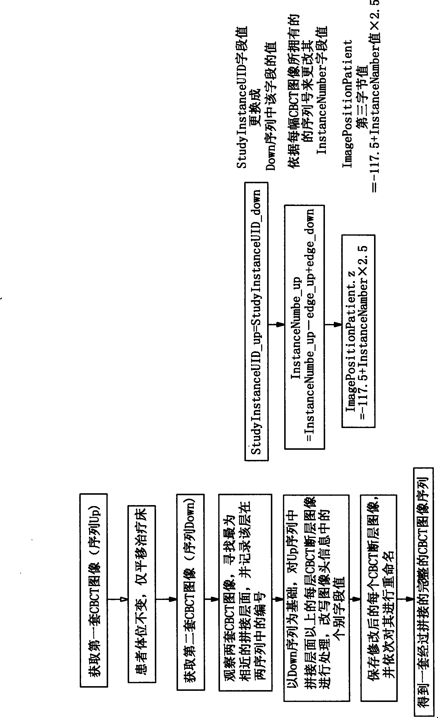
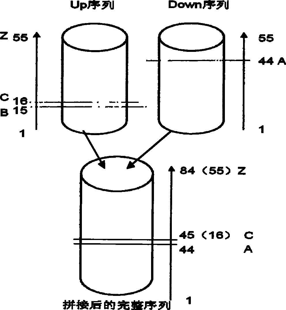
[0037] The steps to obtain CBCT images in segments are as follows:

[0038] 1) Design a CBCT segmented scan plan: Obtain the simulated positioning CT scan image and transfer it to the Varian Eclipse treatment planning system in the accelerator device. Set two isocenters with exactly the same coordinates in the left and right and front and rear directions in the planned CT space. Develop CBCT scan plans P1 and P2 respectively. The length of a single CBCT scan of OBI is 14cm. Therefore, the distance between the two isocenters in the head and foot direction is set within 13cm to ensure that the two CBCT images have sufficient information redundancy;

[0039] 2) CBCT scan: Pass the CBCT plan designed above into the OBI 4D workstation, set up according to the isocenter of plan P1, select the imaging equipment and related parameters according to the scan position, and perform the first CBCT scan. The scan ends And after the online registration is completed, the automatic bed moving param...


PUM


Abstract

The invention provides a method for obtaining an integrated anatomic image through split-jointing subsection cone beam CT images, which comprises the steps as follows: two sections of CBCT scanning plans are respectively set down in plan CT image space; after obtaining a set of CBCT images, a treatment couch is moved horizontally by hand, and CBCT shoot is performed again to obtain adjacent segmented CBCT images; imaging analysis is carried out to the two sections of images respectively, and then the images are processed through image fusion with the plan CT respectively after three-dimensional reconstruction; a splicing lay is selected through the analysis on bone marks after fusion; and two sets of the CBCT images are processed through split-jointing by a computer sequentially to obtaina spliced image of the CBCT images which are obtained through subsection-scanning adjacent parts, thereby enlarging the observing scope of a tumor target area and surrounding normal organs, ensuring the feasibility for setting down a treatment plan by CBCT and the integrity for evaluating dose volume parameters, and having wide clinical application prospect.

Description

1. Technical field [0001] The invention relates to a medical image production technology, in particular to a method for obtaining a complete anatomical image by splicing segmented cone beam CT images. 2. Background technology [0002] The concept of cone-beam CT images: Cone-beam CT, or Cone Beam CT, or CBCT for short, is a type of medical imaging. Similar to ordinary CT images, it is also a tomographic image that reflects the human anatomy and is widely used in the field of tumor radiotherapy. . The image was taken by the "Online Image Collector" (OBI for short) attached to the medical linear accelerator. When a patient receives radiotherapy on this accelerator, the medical technician can control the accelerator head and OBI to rotate concentrically and coplanar around the patient lying on the treatment bed. Make a 360 rotation. Later, the accelerator control terminal stores a set of CBCT image sequences collected by OBI, which can obtain anatomical images within a certain sp...

Claims


Application Information

Patent Timeline
Patent Type & Authority Patents(China)
IPC IPC(8): A61B6/03G06T5/00G06T17/00
Inventor 于金明朱健尹勇刘敏卢洁刘同海陈进琥孙涛白曈张桂芳
Owner AFFILIATED CANCER HOSPITAL OF SHANDONG FIRST MEDICAL UNIV SHANDONG CANCER INST (SHANDONG CANCER HOSPITAL)
Why Eureka
Try Eureka