Abdominal organ segmentation method based on secondary three-dimensional region growth

A three-dimensional area and abdomen technology, applied in the field of medical image processing, can solve the problems of different implementation steps, lack of robustness, lack of unified framework, etc., and achieve the effect of improving robustness and suppressing over-segmentation

Inactive Publication Date: 2009-11-11
XIDIAN UNIV
View PDF0 Cites 32 Cited by
  • Summary
  • Abstract
  • Description
  • Claims
  • Application Information

AI Technical Summary

Problems solved by technology

This method can effectively extract the organs of interest, but because the gray value of the abdominal organs is very similar to the gray value of the surrounding tissue, the result appears to be over-segmented; in addition, the segmentation of each organ of interest in this method is based on Its anatomical position is carried out sequentially ...

Method used


Image

  • Abdominal organ segmentation method based on secondary three-dimensional region growth
  • Abdominal organ segmentation method based on secondary three-dimensional region growth
  • Abdominal organ segmentation method based on secondary three-dimensional region growth

Examples


Embodiment Construction

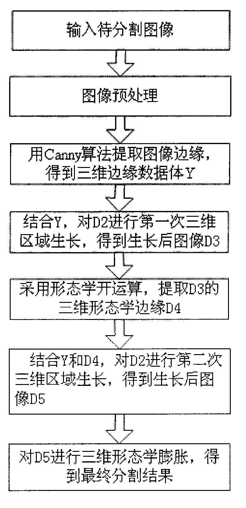
[0023] refer to figure 1 , the specific implementation steps of the present invention are as follows:

[0024] Step 1: Input the image to be segmented.

[0025] Input a set of abdominal CT slices in DICOM format containing the complete liver, spleen and kidney organs. Since the images in DICOM format contain a lot of information that is not related to organ segmentation, in order to reduce the data storage space, only the images related to the image are read here. The information mainly includes the pixel data of each slice and the slice sequence identification number information required in the subsequent processing, so as to obtain a set of slice image data to be divided.

[0026] Step 2: Image preprocessing.

[0027] Preprocessing the sliced ​​image data to be segmented mainly includes: intercepting the body area and removing image noise.

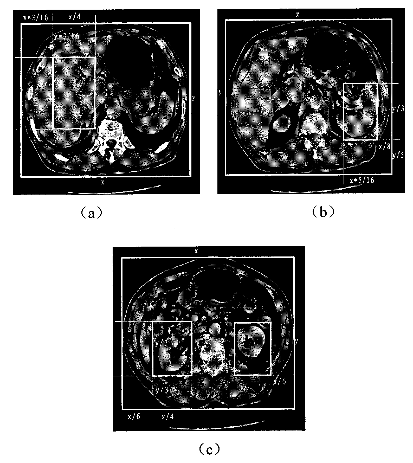
[0028] Capture the body area: Since the abdominal CT image contains a large number of non-body area pixels, in order to further redu...


PUM


Abstract

The invention discloses an abdominal organ segmentation method based on secondary three-dimensional region growth, which belongs to the field of medical image processing. The method comprises the following steps: firstly, combining with apriori knowledge such as anatomical position and gray value distribution of an interested organ to extract original seed points automatically, and combining with image edge extracted by a Canny edge detection algorithm to carry out first three-dimensional region growth of an image; then, extracting the three-dimensional morphologic edge of a segmentation result graph obtained after the first growth; and finally, combining with the extracted three-dimensional morphologic edge and the Canny edge of the original image to carry out second three-dimensional region growth of the original image, and carrying out three-dimensional morphologic expansion of the segmentation result obtained after the second three-dimensional region growth to obtain a final segmentation result of the interested abdominal organ. The abdominal organ segmentation method effectively restrains the phenomenon of oversegmentation existing in the prior three-dimensional region growth method, and can accurately extract an interested organ from an abdominal CT image; therefore, the method can be used for assisting clinical diagnosis.

Description

technical field [0001] The invention belongs to the field of medical image processing and relates to a method for segmenting abdominal organs, which can be used in abdominal CT images to extract interested abdominal organs such as liver, spleen and kidney to assist clinical medical diagnosis. Background technique [0002] The segmentation of abdominal organs has important theoretical value and broad clinical application prospects. Extracting organs of interest from complex backgrounds is the premise and basis for 3D visualization. More importantly, identifying the location and region of the lesion in the organ of interest is helpful for surgery, radiation therapy, etc. The low contrast of medical images, blurred edges, and the uncertainty of different patient organ shapes have made the segmentation of medical images focus on the level of human-computer interaction for a long time. The processing time is long, and the processing results are easily affected by human factors. ...

Claims


Application Information

Patent Timeline
IPC IPC(8): G06T5/00G06T15/20G06T17/00A61B6/03
Inventor 周伟达张莉邹海双武彩丽焦李成
Owner XIDIAN UNIV
Why Eureka
Try Eureka