

Image display device, image display method and image display program
An image display device and image technology, applied in medical images, medical image data management, computer-aided medical procedures, etc., can solve problems that hinder doctors or nurses from diagnosing, and achieve the effect of quickly confirming selected images
- Summary
- Abstract
- Description
- Claims
- Application Information
AI Technical Summary
Problems solved by technology
Method used
Image

Examples
Embodiment approach 1
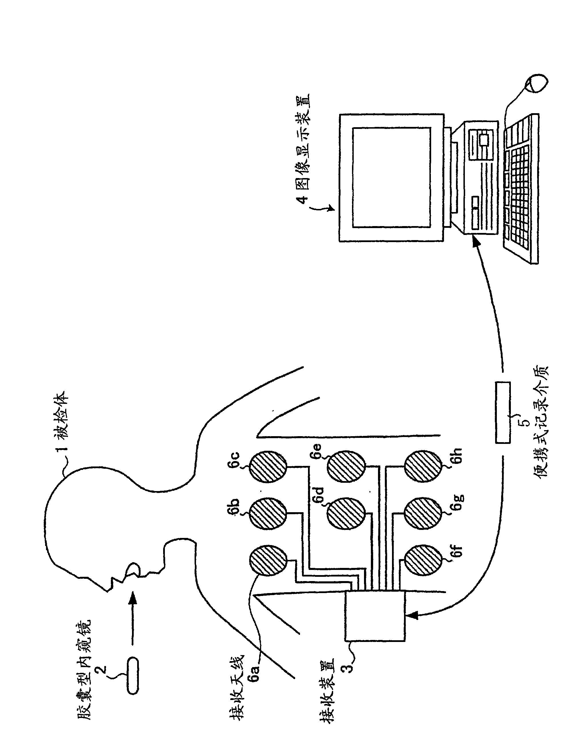
[0078] figure 1 It is a schematic diagram schematically illustrating a configuration example of an in-vivo information acquisition system using the image display device according to Embodiment 1 of the present invention. Such as figure 1 As shown, the system for acquiring information in the subject includes: a capsule endoscope 2 that moves along a passing path in the subject 1 and simultaneously captures images of the subject 1; a receiving device 3 that receives images from the capsule. The image data sent by the capsule endoscope 2; the image display device 4, which displays the images in the subject 1 taken by the capsule endoscope 2 in time series; Information is exchanged between the image display devices 4 .
[0079] The capsule endoscope 2 has an imaging function capable of imaging the inside of the subject, and a wireless communication function for transmitting image data obtained by imaging the inside of the subject to the outside. The capsule endoscope 2 is swall...
Embodiment approach 2
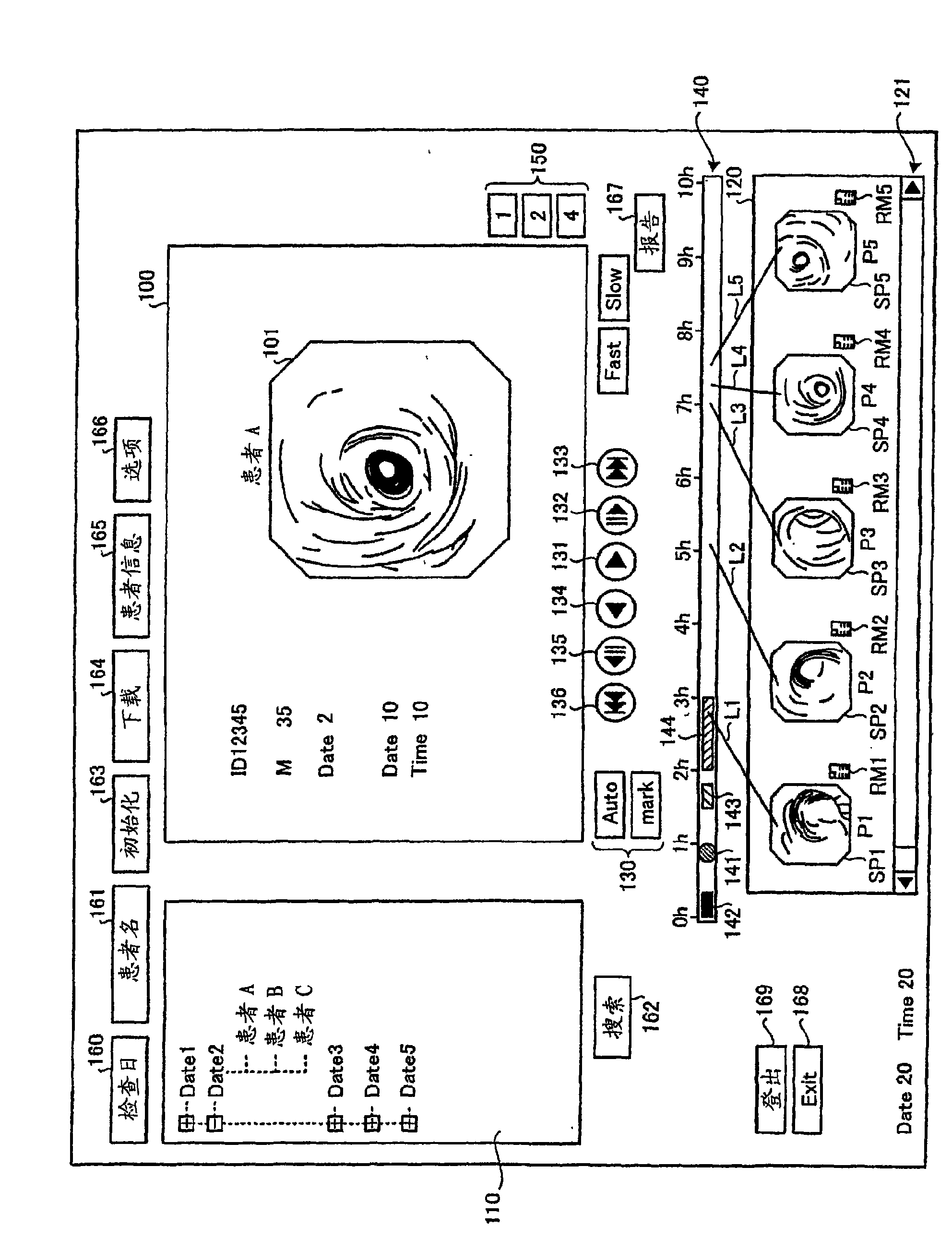
[0180] Next, Embodiment 2 of the present invention will be described in detail. In Embodiment 1 described above, as the indicator indicating the position on the time scale of the time bar 140 corresponding to the shooting date and time of the selected image, a sub-image such as a thumbnail image corresponding to the selected image is displayed on the time scale. However, in Embodiment 2, an in-vivo schematic image schematically showing the passing path of the capsule endoscope 2 in the subject 1 is displayed, and the sub-image is displayed on top of the in-vivo schematic image. Instead of the indicator that associates the secondary image with the position on the time scale, the indicator associated with the position of the time scale.
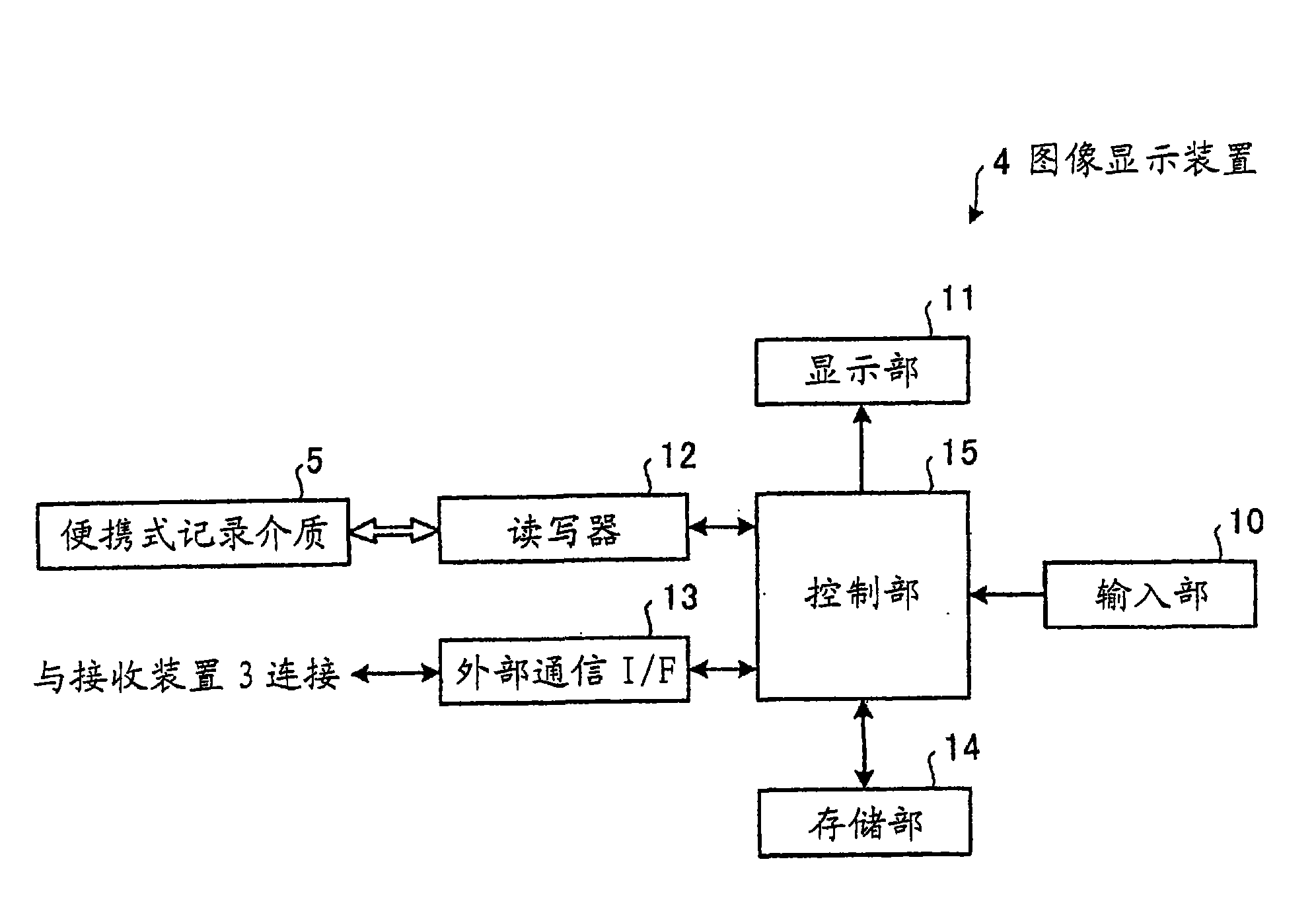
[0181] Figure 18 It is a block diagram schematically showing a configuration example of an image display device according to Embodiment 2 of the present invention. This image display device 21 is provided with a control unit 22 instead of th...
Embodiment approach 3
[0197] Next, an image display device according to Embodiment 3 of the present invention will be described with reference to the drawings. The image display device according to Embodiment 3 displays an image selected by a user in a predetermined selected image display area among images sequentially displayed in a predetermined image display area. In addition, in description of drawings, the same code|symbol is attached|subjected to the same part.
[0198] Figure 20 It is a schematic diagram showing the overall configuration of the in-vivo information acquisition system in the third embodiment. Such as Figure 20 As shown, the in-body information acquisition system in Embodiment 3 includes: a receiving device 302 having a receiving function; and a capsule endoscope 303 that is introduced into the body of the subject 1 to photograph The images in the body cavity are sent to the receiving device 302 as data. In addition, the in-body information acquisition system includes: an...
PUM
 Abstract
Description
Claims
Application Information