Image display device, image display method and image display program

An image display device and image display technology, applied in the direction of surgery, etc., can solve the problems that hinder the diagnosis of doctors or nurses, and achieve the effect of quickly confirming the selected image

Inactive Publication Date: 2009-11-25
OLYMPUS CORP
View PDF12 Cites 2 Cited by
  • Summary
  • Abstract
  • Description
  • Claims
  • Application Information

AI Technical Summary

Problems solved by technology

However, when a doctor or a nurse checks a selected image on a monitor, it is necessary to perform complicated processing of retrieving a desired selected image from a plurality of stored images and displaying the retrieved selected image on the monitor, which sometimes hinders the doctor. or a nurse to make a quick diagnosis

Method used


Image

  • Image display device, image display method and image display program
  • Image display device, image display method and image display program
  • Image display device, image display method and image display program

Examples


Embodiment approach 1

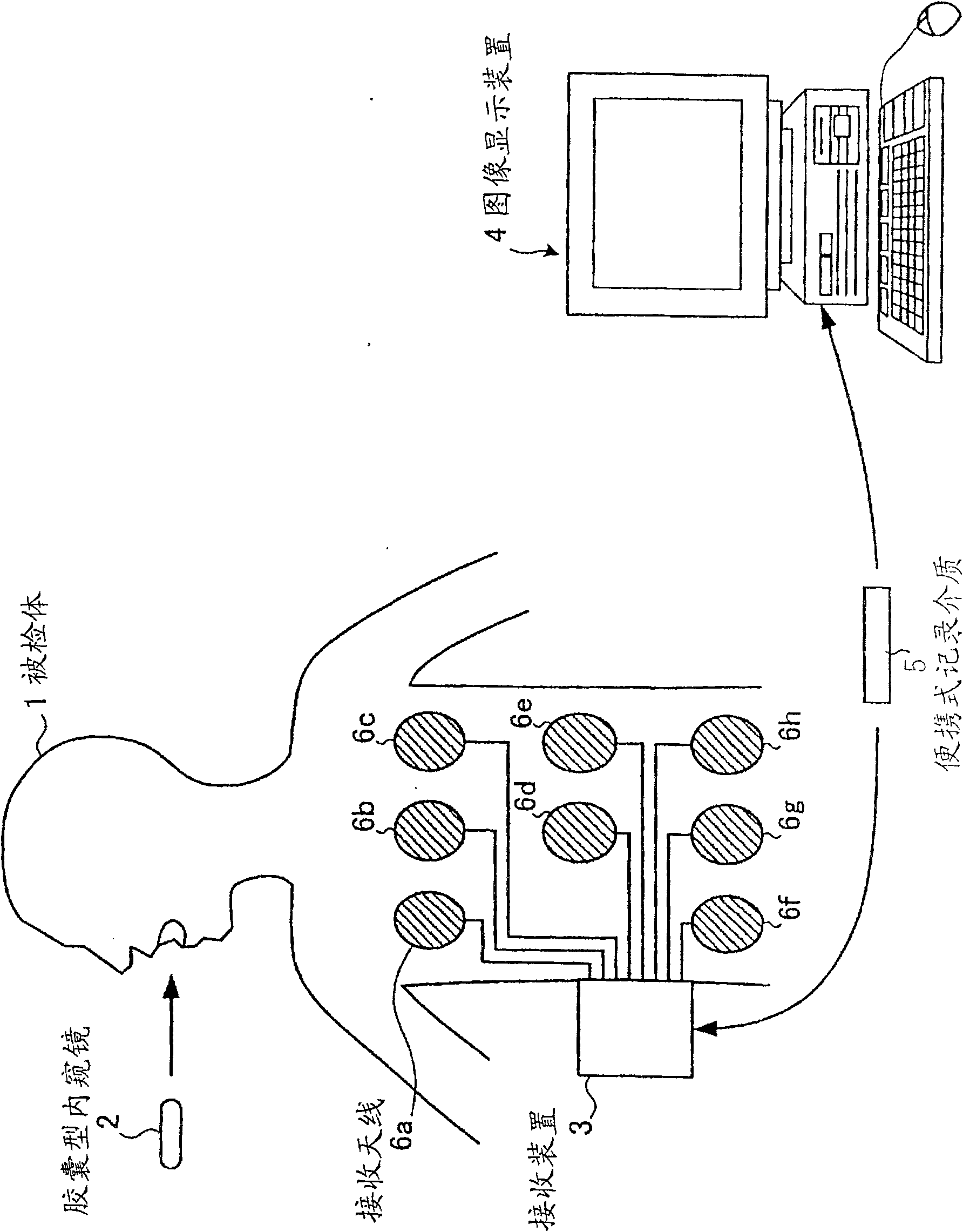
[0054] figure 1 It is a schematic diagram schematically illustrating a configuration example of an in-vivo information acquisition system using the image display device according to Embodiment 1 of the present invention. Such as figure 1 As shown, the system for acquiring information in the subject includes: a capsule endoscope 2 that moves along a passing path in the subject 1 and simultaneously captures images of the subject 1; a receiving device 3 that receives images from the capsule. The image data sent by the capsule endoscope 2; the image display device 4, which displays the images in the subject 1 taken by the capsule endoscope 2 in time series; Information is exchanged between the image display devices 4 .

[0055] The capsule endoscope 2 has an imaging function capable of imaging the inside of the subject, and a wireless communication function for transmitting image data obtained by imaging the inside of the subject to the outside. The capsule endoscope 2 is swall...

Embodiment approach 2

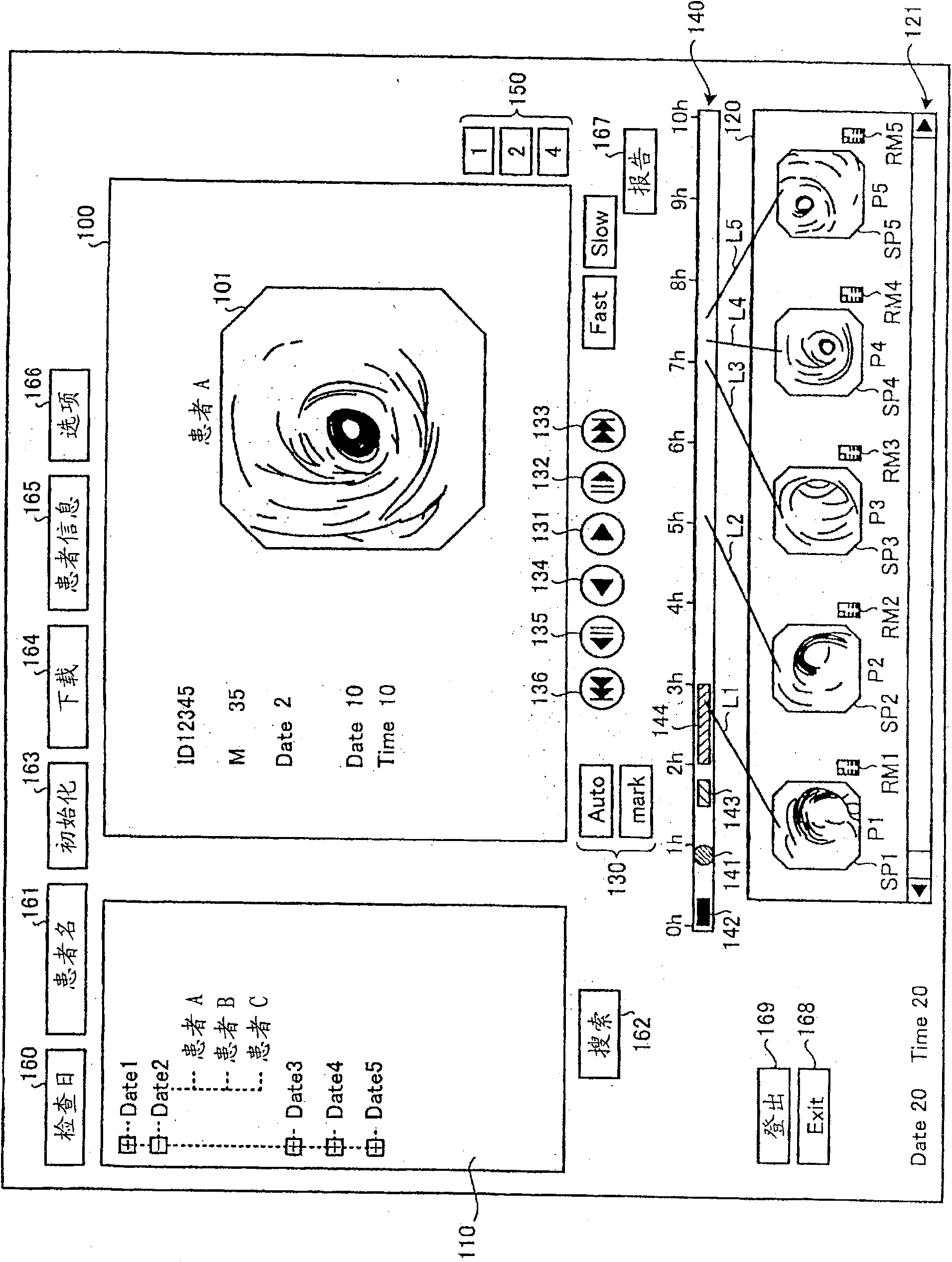
[0156] Next, Embodiment 2 of the present invention will be described in detail. In Embodiment 1 described above, as the indicator indicating the position on the time scale of the time bar 140 corresponding to the shooting date and time of the selected image, a sub-image such as a thumbnail image corresponding to the selected image is displayed on the time scale. However, in Embodiment 2, an in-vivo schematic image schematically showing the passing path of the capsule endoscope 2 in the subject 1 is displayed, and the sub-image is displayed on top of the in-vivo schematic image. Instead of the indicator that associates the secondary image with the position on the time scale, the indicator associated with the position of the time scale.

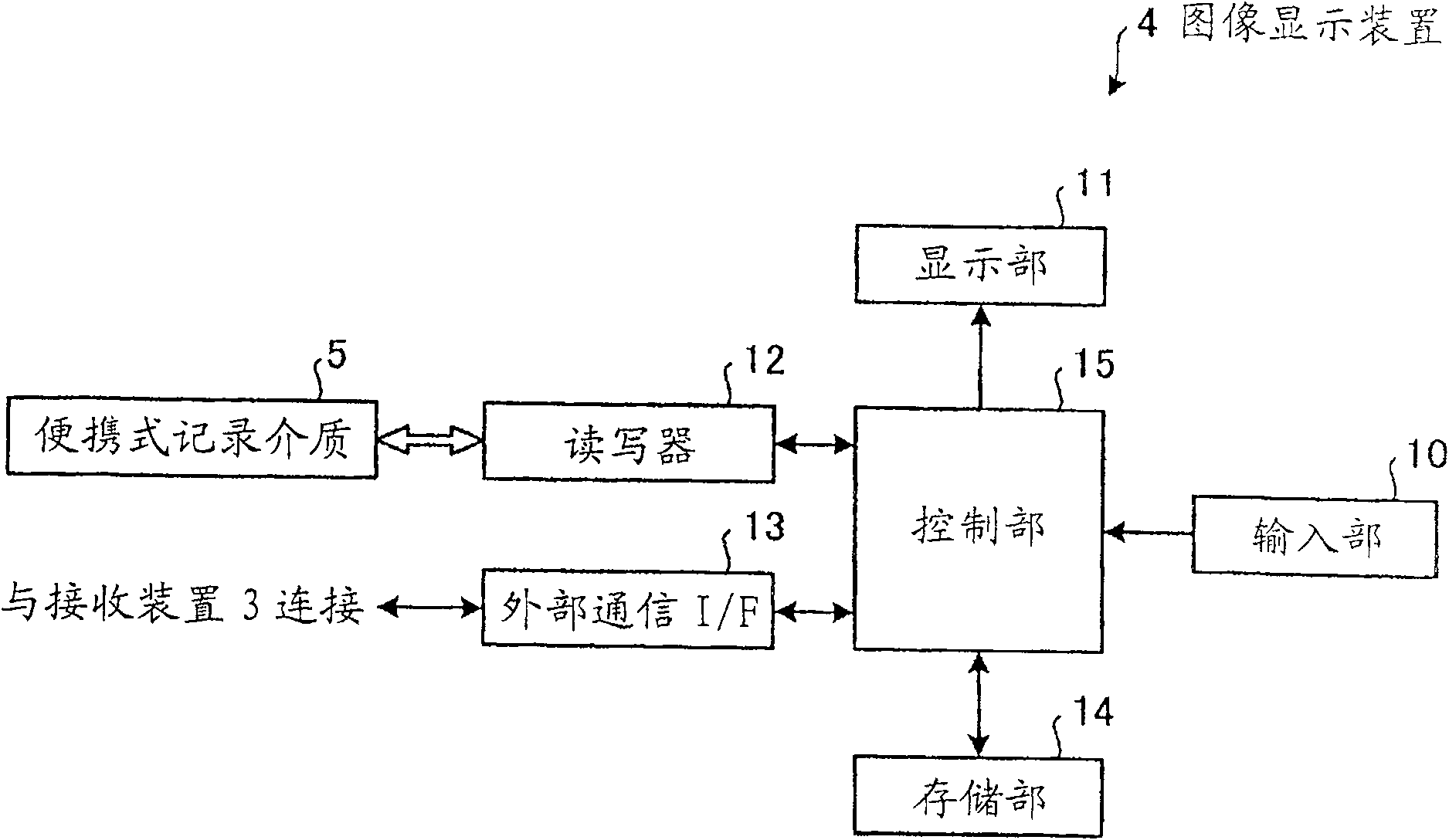
[0157] Figure 18 It is a block diagram schematically showing a configuration example of an image display device according to Embodiment 2 of the present invention. This image display device 21 is provided with a control unit 22 instead of th...

Embodiment approach 3

[0173] Next, an image display device according to Embodiment 3 of the present invention will be described with reference to the drawings. The image display device according to Embodiment 3 displays an image selected by a user in a predetermined selected image display area among images sequentially displayed in a predetermined image display area. In addition, in description of drawings, the same code|symbol is attached|subjected to the same part.

[0174] Figure 20 It is a schematic diagram showing the overall configuration of the in-vivo information acquisition system in the third embodiment. Such as Figure 20 As shown, the in-body information acquisition system in Embodiment 3 includes: a receiving device 302 having a receiving function; and a capsule endoscope 303 that is introduced into the body of the subject 1 to photograph The images in the body cavity are sent to the receiving device 302 as data. In addition, the in-body information acquisition system includes: an...


PUM


Abstract

The present invention provides an image display device capable of easily estimating the shooting position of a selected image from a display screen. The image display device of the present invention includes a display unit and a control unit that display a series of images obtained by photographing a desired subject in time series. The control section controls the display section to display a time bar to which a time scale representing the shooting period of the series of images is added, and controls the display section to display a time bar indicating and selecting the image selected from the series of images. An indication mark of a position on the time scale corresponding to the shooting moment of the desired image.

Description

technical field [0001] The present invention relates to an image display device, an image display method, and an image display program that display a sequence of images obtained by photographing a desired subject in time series. Background technique [0002] In recent years, in the field of endoscopy, capsule-type endoscopes equipped with imaging functions and wireless communication functions have been proposed as swallowing-type endoscopes. An acquisition system acquires image data inside a subject using the capsule endoscope. This capsule endoscope plays a role in that, after being swallowed from the mouth of the subject for observation (examination) until it is naturally discharged from the subject, in the subject such as the stomach or The inside of an internal organ such as the small intestine moves along with the peristaltic motion of the internal organ, and at the same time, the inside of the subject is imaged at predetermined intervals, for example, at intervals of ...

Claims


Application Information

Patent Timeline
Patent Type & Authority Patents(China)
IPC IPC(8): A61B1/00G06F3/048G06F3/0484
Inventor 藤田学平川克己
Owner OLYMPUS CORP
Why Eureka
Try Eureka