A cell-mediated immune response assay and kits therefor
A kit, immune effect technology, applied in the determination/inspection of microorganisms, material testing products, biological testing, etc., can solve problems such as reducing sensitivity
- Summary
- Abstract
- Description
- Claims
- Application Information
AI Technical Summary
Problems solved by technology
Method used
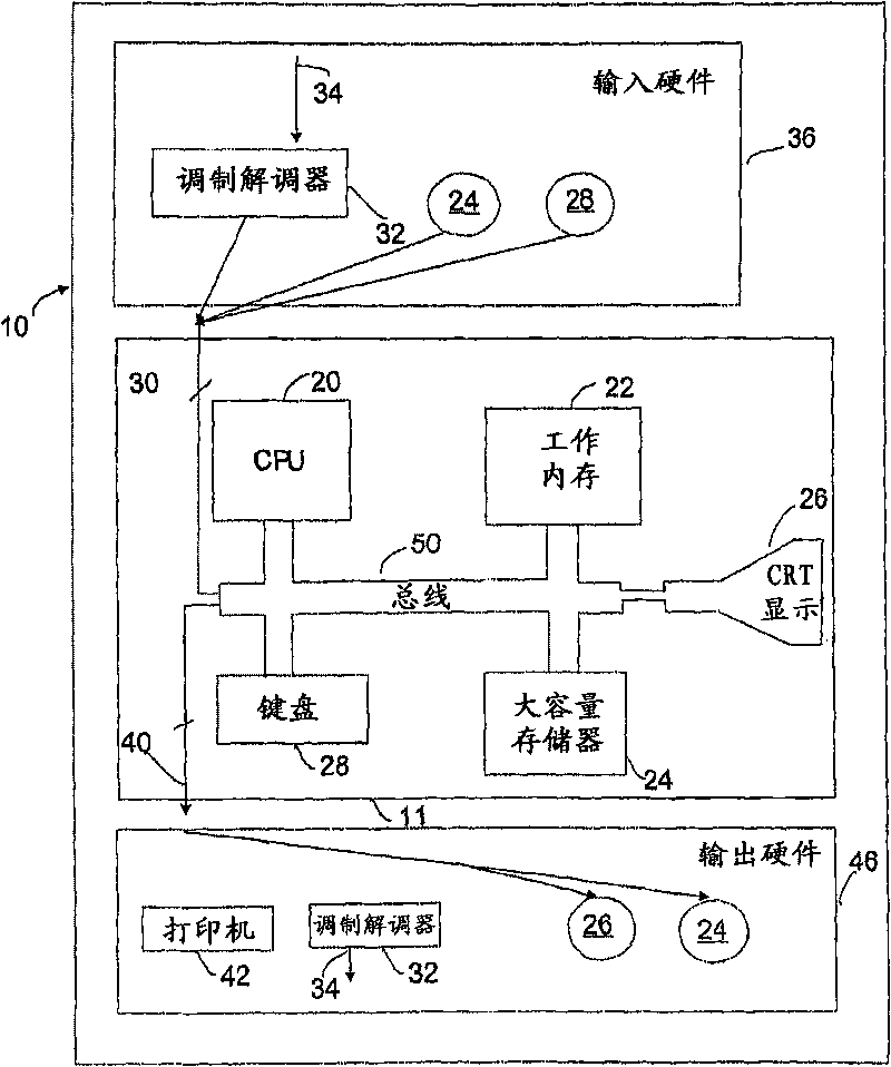
Image

Examples
Embodiment 1
[0130] Detection of immune effector molecules in small volume samples incubated with antigen
[0131] Whole blood from healthy donors (4 donors) was collected into 9 mL Vacuette Li-heparin tubes with approximately 6.6 mm diameter cylinders and U-shaped bases. Aliquots of 0.1, 0.2, 0.3, 0.4 and 0.5 mL of blood (no additives) were stimulated in Vacuette micro collection tubes. Blood was stimulated with tetanus toxoid and phytohemagglutinin-P (mitogen). The volume of antigen added to each tube is proportional to the volume of blood, eg, 0.1 mL of blood is stimulated with 0.01 mL of antigen and 0.4 mL of blood is stimulated with 0.04 mL of antigen.
[0132] The IFN-γ responses generated in small volumes of blood were compared to those generated in 13 / 75 Vacuette blood collection tubes (control) using 1 mL of blood.
[0133] Blood was incubated with antigen at 37°C for 16 to 24 hours before removal of plasma for IFN-γ detection (QuantiFERON-TB Gold ELISA (QuantiFERON-TB Gold ELIS...
Embodiment 2
[0137] Detection of Immune Effector Molecules in Small Volumes of Whole Blood Incubated in Containers with Different Internal Shapes
[0138] Heparinized blood was dispensed into 3x5 mL aliquots in polypropylene tubes. Antigens, either human cytomegalovirus (CMV) or tetanus toxoid (Tetanus), are added to the blood at appropriate concentrations. Each tube was mixed thoroughly so that the blood (50 μl) was dispensed into various containers where the blood volume took different heights. Specifically, PCR tubes (conical, height 10 mm, maximum diameter 5 mm, U-shaped base), micro-collectors (cylindrical, 6.5 mm diameter, U-shaped base), 96-well plates (cylindrical, 6.5 mm diameter, flat bottom) , 48-well plate (cylindrical, 11 mm in diameter, flat bottom). Containers were incubated at 37°C for 20 hours before removal of plasma (20 μl) for testing by QFT-ELISA using labeled antibodies to detect IFN-γ production. In a control experiment, 1 mL of blood was incubated in a cylindrica...
Embodiment 3
[0140] Detection of immune effector molecules in peripheral capillary blood
[0141] Capillary blood (150 μl) was collected by finger prick into lithium heparin micro-collection tubes. Transfer small volumes (50 μl) to three PCR tubes. The PCR tubes are conical with a cylindrical upper portion that tapers from 10mm to a conical base. Antigens for CMV or Tetanus toxoid (Tetanus) or no antigen (Nil) were added to the tubes, which were incubated at 37°C for 20 hours. Thereafter, plasma (20 μl) was removed and tested using QFT-ELISA. As shown in Figure 5, the positive CMV control subject (TR) samples produced a significant signal compared to the negative controls, indicating that the assay can be performed in volumes as small as 50 μl of capillary blood.
PUM
 Abstract
Description
Claims
Application Information
 - R&D Engineer
- R&D Manager
- IP Professional
- Industry Leading Data Capabilities
- Powerful AI technology
- Patent DNA Extraction
Browse by: Latest US Patents, China's latest patents, Technical Efficacy Thesaurus, Application Domain, Technology Topic.
© 2024 PatSnap. All rights reserved.Legal|Privacy policy|Modern Slavery Act Transparency Statement|Sitemap