
Dental device and method for linking physical and digital data for diagnostic, treatment planning, patient education, communication, manufacturing, and data transfer purposes

A dental, patient technique, applied in the direction of instruments for radiology, diagnostics, dentistry, etc., capable of solving problems such as ineffectiveness of attempts to match areas and points from model scans

Inactive Publication Date: 2011-10-12
IBUR
View PDF0 Cites 9 Cited by
  • Summary
  • Abstract
  • Description
  • Claims
  • Application Information

AI Technical Summary

Problems solved by technology

This procedure is problematic if the CT image has scatter, as attempts to match regions and points from model scans may not work

Method used


Image

  • Dental device and method for linking physical and digital data for diagnostic, treatment planning, patient education, communication, manufacturing, and data transfer purposes
  • Dental device and method for linking physical and digital data for diagnostic, treatment planning, patient education, communication, manufacturing, and data transfer purposes
  • Dental device and method for linking physical and digital data for diagnostic, treatment planning, patient education, communication, manufacturing, and data transfer purposes

Examples


Embodiment Construction

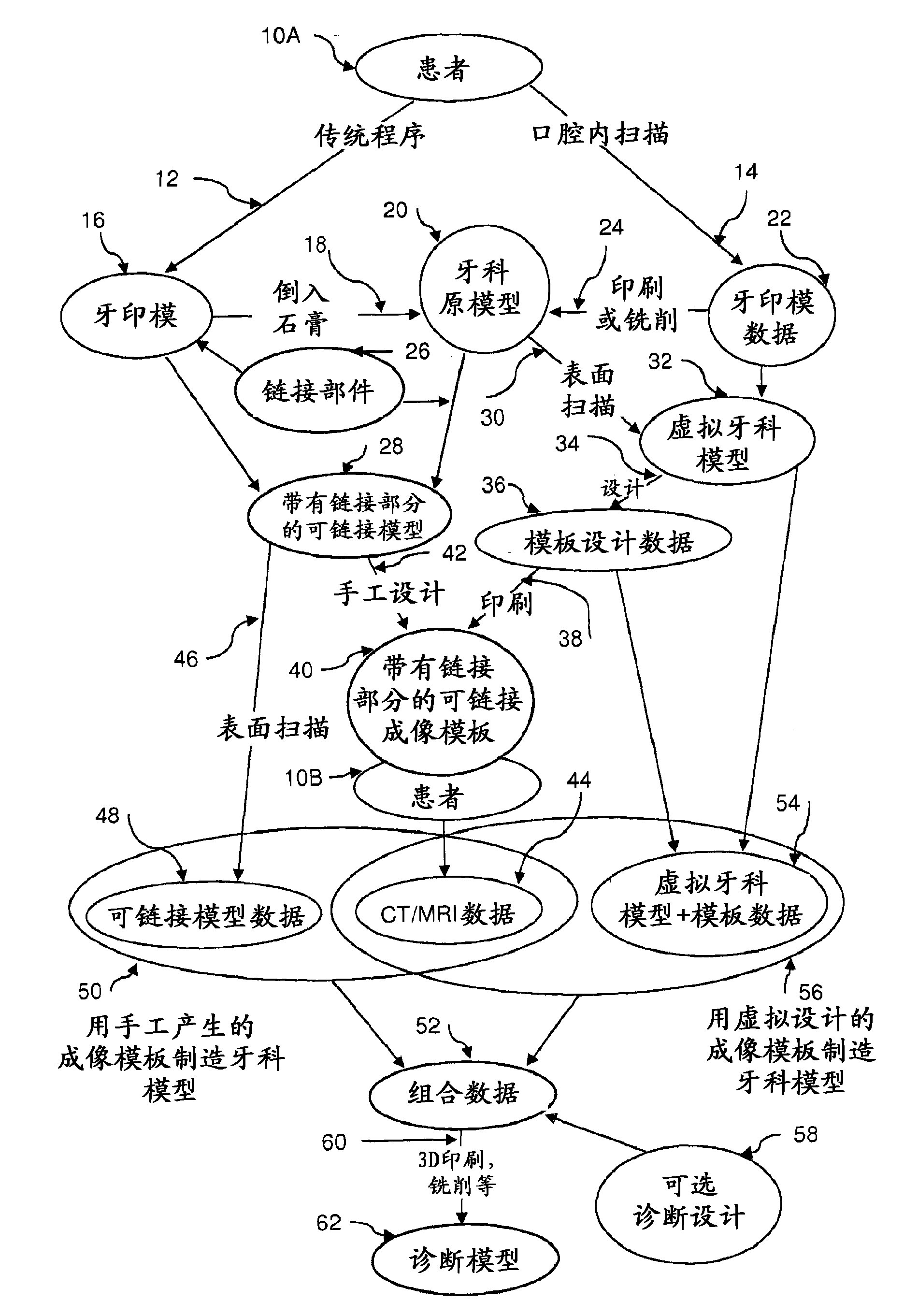
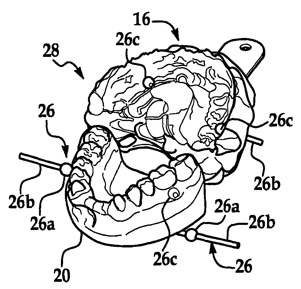
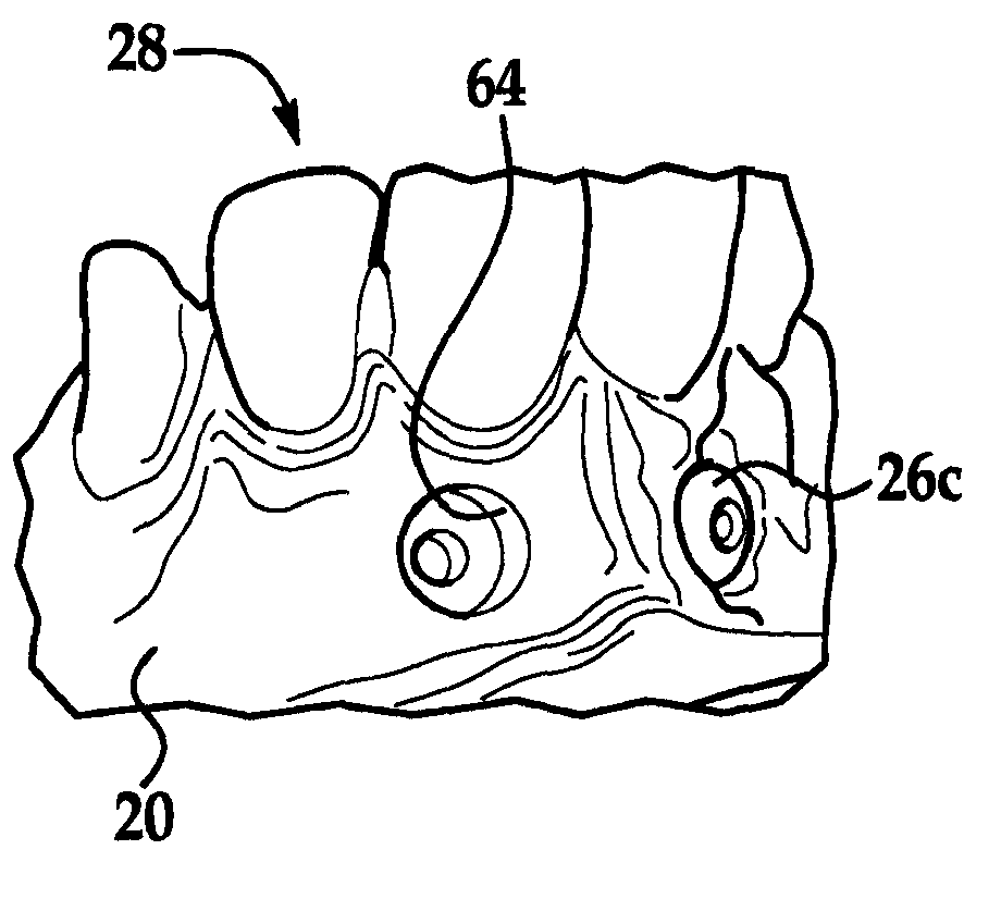
[0038] now refer to figure 1 , a simplified schematic showing an information linking mechanism with linking components begins with a particular patient undergoing a conventional procedure 12 or an intraoral scan 14 at a first point in time 10A. The traditional procedure 12 includes a dental impression 16 , pouring plaster 18 to create a dental master model 20 . The intraoral scan 14 includes dental impression data 22 , which is printed or milled 24 to produce a dental master model 20 . A linking component 26 may be associated with the dental impression 16 and the dental master model 20 to define a linkable model 28 with linked parts. The linking component 26 may be used to generate an identifiable link of imaging scan data common to both the surface scan data file and the tomographic scan data file. The dental master model 20 may be surface scanned 30 to generate a surface scan data file, or the dental impression data may be inverted to provide a virtual dental model 32 . T...


PUM


Abstract

A dental device and / or process include at least one scaled and shaped linking component (26) to be supported by a dental model (28) or an imaging template (40). The process includes scaling, aligning, and orienting data (50, 56) from different data acquisition sources (44, 48, 54) with the scaled and shaped linking component (26), and combining the data (50, 56) from different data acquisition sources into a master data file (52). A method of making a diagnostic model (62) can include virtually designing an imaging template (36) including at least one linking component (26) made at least partially of a radio opaque material, and three-dimensionally printing the virtually designed template (40).; The diagnostic model (62) can include at least one of an exposed bone structure portion (70a, 76a), a removable gum tissue portion (72, 76b), a removable bone structure portion (76c), a visualization portion illustrating a root (76d), bone density (76f), an internal bone structure (76g), a nerve channel (76h), a nerve ending (76i), a sinus cavity (76j), a blood vessel (76k), an artery (761), and diagnostic teeth (76r).

Description

[0001] related invention [0002] This application claims prior U.S. Provisional Patents Serial No. 61 / 115,874 filed November 18, 2008 and Serial No. 61 / 270,942 filed July 15, 2009 pursuant to 35 U.S.C. § 119(e) priority of the application, both of which are hereby incorporated by reference in their entirety. technical field [0003] The field of the invention relates to dental devices and procedures from imaging and other sources of information associated with various data sets in physical and / or digital Datasets of collected information linked into a comprehensive digital format for virtual design / illustration and manufacture of image scan templates, surgical guides, implants, crowns, bridges, and / or with the aid of treatment determined appropriate for a particular patient A template for optional diagnostic components useful for the process. Background technique [0004] The physical master dental model may relate to medical, dental trauma edentulous, partially edentulou...

Claims


Application Information

Patent Timeline
Patent Type & Authority Applications(China)
IPC IPC(8): A61C19/04A61B6/14A61B6/51
CPCA61C9/00A61C9/0053A61B6/506G16H50/50B33Y80/00A61C9/004A61C13/34
Inventor R·C·格罗斯库特S·U·格罗斯库特
Owner IBUR
Learn More