
Mr imaging with cest contrast enhancement

A technique of imaging sequences, images, applied in the field of computer programs, which can solve problems such as negative effects on patient comfort

Inactive Publication Date: 2011-11-23
KONINK PHILIPS ELECTRONICS NV
View PDF4 Cites 13 Cited by
  • Summary
  • Abstract
  • Description
  • Claims
  • Application Information

AI Technical Summary

Problems solved by technology

Imaging times can easily exceed ten minutes, which has a negative impact on patient comfort

Method used


Image

  • Mr imaging with cest contrast enhancement
  • Mr imaging with cest contrast enhancement
  • Mr imaging with cest contrast enhancement

Examples


Embodiment Construction

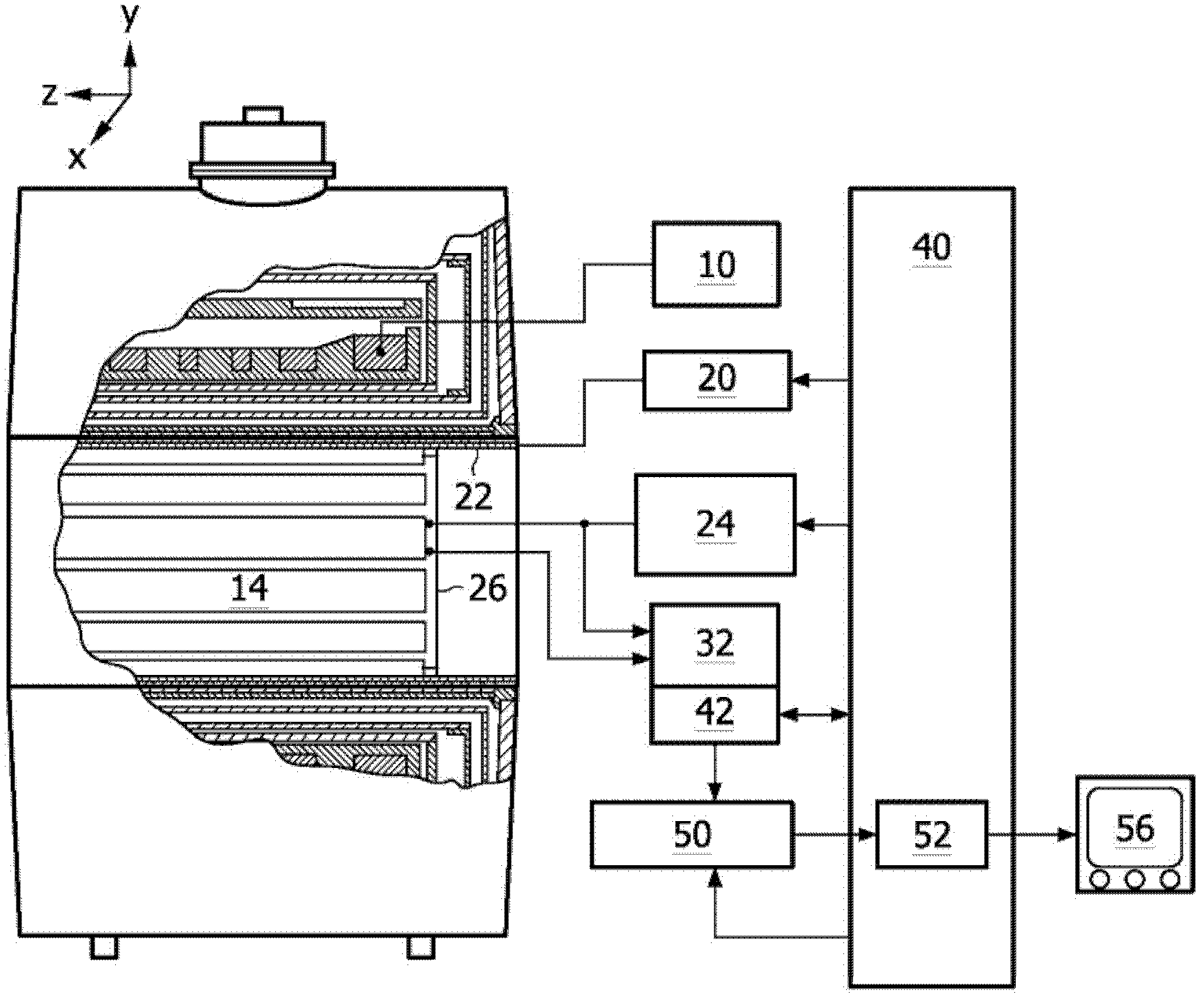
[0041] refer to figure 1 , the main magnetic field controller 10 controls the superconducting or resistive main magnet 12 to generate a substantially uniform, temporally constant main magnetic field along the z-axis through the examination volume 14 .

[0042] Magnetic resonance generation and manipulation systems apply a series of RF pulses and switched magnetic field gradients to flip or excite nuclear magnetic spins, induce magnetic resonance, refocus magnetic resonance, manipulate magnetic resonance, spatially encode or otherwise encode magnetic resonance, such that Spin saturation etc. to perform MR imaging.

[0043]More specifically, gradient pulse amplifier 20 applies current pulses to selected ones of pairs of whole body gradient coils 22 to generate magnetic field gradients along the x, y, and z axes of examination volume 14 . A digital RF frequency transmitter 24 transmits RF pulses or bursts to a whole body RF coil 26 for transmitting RF pulses into the examination...


PUM


Abstract

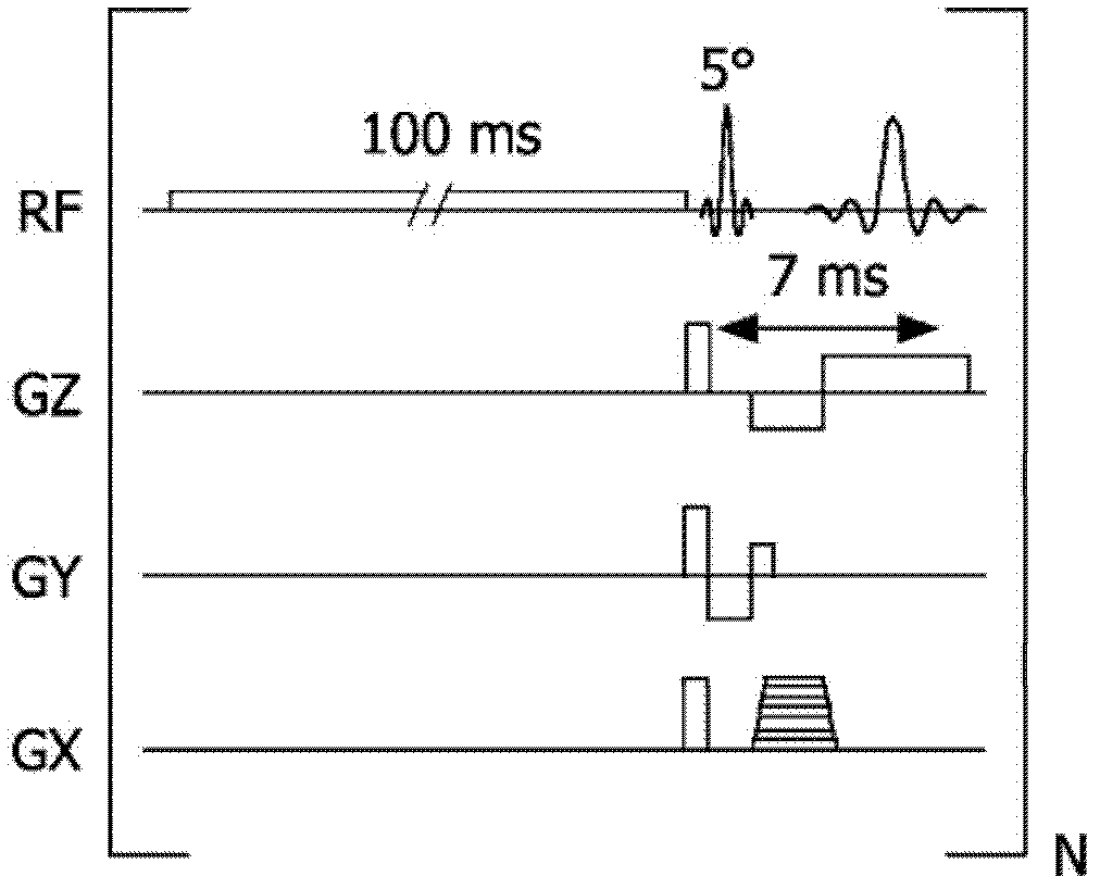
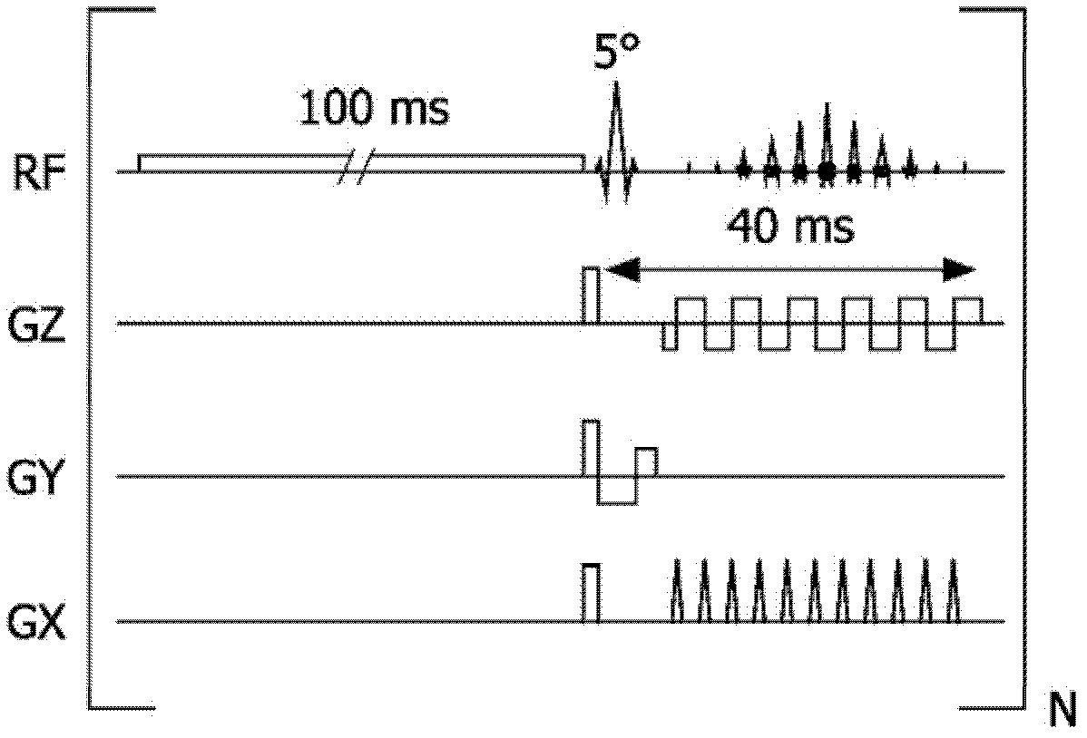
The invention relates to a method of MR imaging of at least a portion of a body of a patient placed in an examination volume of an MR device. The object of the invention is to improve CEST contrast enhanced imaging. The method of the invention comprises the following steps: a) saturation of nuclear magnetization of exchangeable protons of a CEST contrast agent administered to the patient by subjecting the portion of the body to at least one frequency-selective saturation RF pulse matched to the MR frequency of exchangeable protons of the CEST contrast agent, wherein the saturation period, i.e. the duration of the frequency-selective saturation RF pulse, is shorter than the time required for saturation to build up a full CEST contrast enhancement effect when starting from zero saturation; b) generating at least one MR signal of water protons of the body by subjecting the portion of the body to an MR imaging sequence comprising at least one RF pulse and switched magnetic field gradients; c) acquiring and sampling the at least one MR signal from the body; d) repeating steps a) to c) a number of times under variation of parameters of the MR imaging sequence, wherein MR signals are acquired and sampled during a saturation build-up period, i.e. before a steady state of the CEST effect is achieved; e) reconstructing a proton-density weighted, CEST contrast-enhanced MR image from the acquired and sampled MR signals.

Description

technical field [0001] The present invention relates to the field of magnetic resonance (MR) imaging. The invention relates to a method for MR imaging of at least a part of a patient's body placed in an examination volume of an MR apparatus. The invention also relates to an MR device and a computer program to be run on the MR device. Background technique [0002] Image-forming MR methods that exploit the interaction between magnetic fields and nuclear spins to form two- or three-dimensional images are currently in widespread use, especially in the field of medical diagnostics, as they are superior to other imaging in many ways for soft tissue imaging method, does not require ionizing radiation and is usually noninvasive. [0003] According to the general MR method, the body of the patient to be examined is arranged in a strong homogeneous magnetic field, the direction of which simultaneously defines the axis (usually the z-axis) of the coordinate system on which the measur...

Claims


Application Information

Patent Timeline
Patent Type & Authority Applications(China)
IPC IPC(8): G01R33/28
CPCG01R33/5616G01R33/5601
Inventor J·A·皮克马特S·朗戈埃斯H·格吕尔D·布丁斯基R·M·J·N·拉默里奇斯J·库普
Owner KONINK PHILIPS ELECTRONICS NV
Learn More