Method and apparatus for obtaining and processing ballistocardiograph data

A technology that processes data and shocks the heart. It is used in medical science, telemetry patient monitoring, sensors, etc., and can solve problems such as expensive equipment.

Inactive Publication Date: 2012-05-16
HEART FORCE MEDICAL
View PDF7 Cites 19 Cited by
  • Summary
  • Abstract
  • Description
  • Claims
  • Application Information

AI Technical Summary

Problems solved by technology

Echocardiography provides images of parts of the heart and can provide understandable information about the structure and function of that heart, but requires expensive equipment and specialized operators

Method used


Image

  • Method and apparatus for obtaining and processing ballistocardiograph data
  • Method and apparatus for obtaining and processing ballistocardiograph data
  • Method and apparatus for obtaining and processing ballistocardiograph data

Examples


Embodiment 1

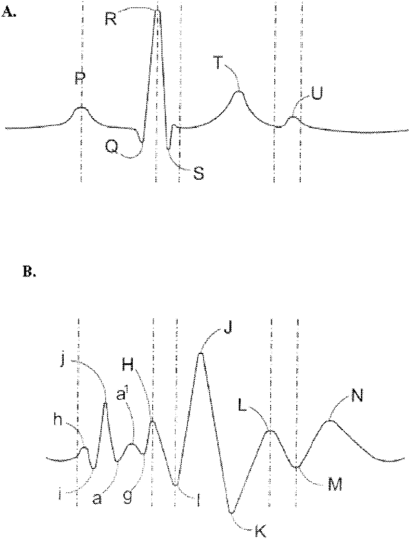
[0136] The following examples relate to specific methods for analyzing ballistocardiography (BCG) data. Figure 6An embodiment corresponding to a simultaneous electrocardiogram-ballistocardiograph (ECG-BCG) waveform set 200 including interpretations from a triaxial accelerometer is shown in . The ECG-BCG waveform set is a visual representation of ECG signal data 210 and BCG signal data 202, 204, 206 captured using the sensor device 10'. Because the detection of ECG and BCG signals by the sensor device begins simultaneously in response to the "start" command, the ECG-BCG waveform set 200 is automatically synchronized in time. As shown, the ballistocardiogram includes three separate waveforms corresponding to different axes of the accelerometer. These waveforms are identified by showing the x-axis waveform 202 as a dashed line, the y-axis waveform 204 as a thin line, and the z-axis waveform 206 as a thick line. When using the sensor device 10, no ECG data is provided.

[0137...

Embodiment 2

[0184] refer to Figure 16 , shows a method of determining an index related to the physiological condition of the subject 52 . At step 54, a derived numerical waveform is provided. A thrust summation is then determined between the first annotation and the second annotation, which is located using predefined rules and divided by the length of time between the first annotation and the second annotation to provide respectively as in step 56 and the index indicated at 58. The first annotation and the second annotation are located using a rule set including rules for locating each cardiac event on the derived numerical waveform. At step 60 , the index is compared to one of the overall standard index or the individual correlation index and a report is output, as indicated at step 62 .

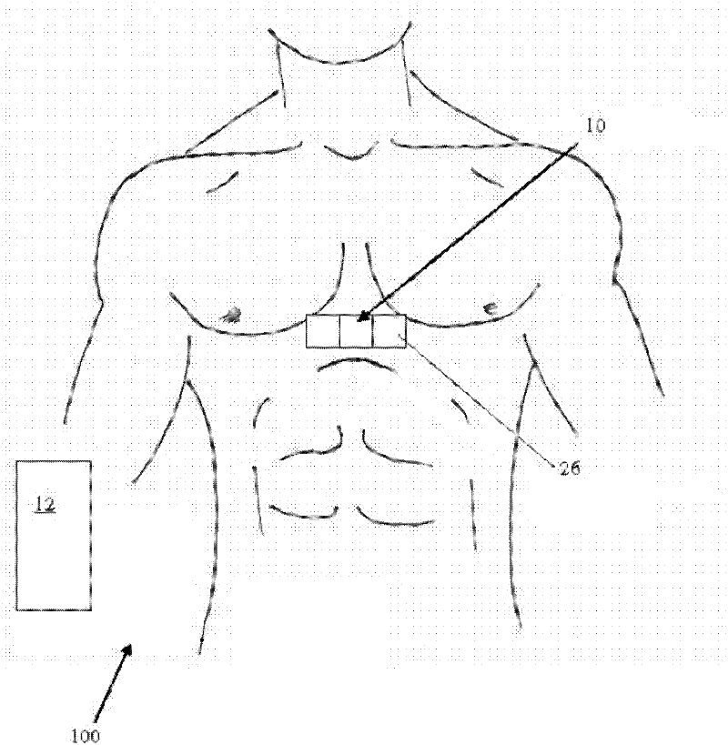
[0185] In operation, the sensor device 10 is coupled to the subject's sternum using adhesive and BCG signals are detected therefrom. When coupled to the chest, orienting the sensor device such th...

Embodiment 3

[0192] Other time intervals of interest may be defined for the processing of the ballistocardiogram data. For example, ballistocardiogram data during different periods of the cardiac cycle, which are known in the art for each of the heart valves assigned to them, may be known to suffer from impairment of the function of the heart valve Individuals are processed and can be compared to data from individuals with standard valve function. Comparison of data within these specific time windows will assist the physician in the diagnosis and measurement of the severity of the valve dysfunction.


PUM


Abstract

A method and apparatus are provided for obtaining and processing ballistocardiograph data to determine a physiological condition of a subject. Ballistocardiograph data indicative of heart motion of the subject measured along a plurality of spatial axes by a sensor device which may comprise a three-axis accelerometer. The ballistocardiograph data is processed to determine processed data indicative of heart motion of the subject. Indications of physiological condition are determined based at least in part on the processed data. Processing may comprise aggregation of multidimensional data, determining magnitude of heart motion and derivative thereof, determining a thrust summation, determining an index value, outputting a report based on an index value, etc. Processing may be informed by operator input, such as a time window of interest or indications of interest.

Description

field of invention [0001] The present invention relates generally to techniques for determining the physiological condition of a subject and, in particular, to methods and apparatus for obtaining and processing ballistocardiogram data. Background technique [0002] Cardiovascular disease is one of the leading causes of death in the Western world, and its incidence is expected to increase in the coming years as this 'baby boom' takes shape. As a result, technologies that improve or aid in the assessment and / or detection of cardiovascular disease will become increasingly important in patient monitoring and overall care. [0003] Currently, devices such as electrocardiogram (ECG) monitors and echocardiography are used in the identification and assessment of cardiovascular disease. The ECG provides a fairly quick electronic assessment of the heart, but does not provide any information related to contractility. Echocardiography provides images of parts of the heart and can prov...

Claims


Application Information

Patent Timeline
Patent Type & Authority Applications(China)
IPC IPC(8): A61B5/02A61B19/00A61B5/11
CPCA61B5/02028A61B5/7239A61B5/0002A61B5/1102A61B5/029A61B5/7282A61B2562/0219A61B5/726A61B5/7278
Inventor 爱德华兹·巴瑟格雷姆·杨斯詹姆斯·亚历山大·伯恩斯
Owner HEART FORCE MEDICAL
Why Eureka
Try Eureka