Intraoperative CT (Computed Tomography) image beam hardening artifact correction method and device

A CT image and beam hardening technology, applied in image enhancement, image data processing, instruments, etc., can solve problems such as artifacts, achieve the effect of improving correction efficiency, speeding up image processing speed, and reducing data processing volume

Active Publication Date: 2014-11-26
SHENZHEN ANKE HIGH TECH CO LTD
View PDF3 Cites 4 Cited by
  • Summary
  • Abstract
  • Description
  • Claims
  • Application Information

AI Technical Summary

Problems solved by technology

[0005] In view of the above deficiencies in the prior art, the purpose of the present invention is to provide a method and device for correcting intraoperative CT image beam hardening artifacts, aiming to solve the artifact problem caused by the intraoperative CT beam hardening phenomenon in the prior art

Method used


Image

  • Intraoperative CT (Computed Tomography) image beam hardening artifact correction method and device
  • Intraoperative CT (Computed Tomography) image beam hardening artifact correction method and device
  • Intraoperative CT (Computed Tomography) image beam hardening artifact correction method and device

Examples


Embodiment Construction

[0054] The present invention provides a method and device for correcting intraoperative CT image beam hardening artifacts. In order to make the purpose, technical solution and effect of the present invention clearer and clearer, the present invention will be further described in detail below. It should be understood that the specific embodiments described here are only used to explain the present invention, not to limit the present invention.

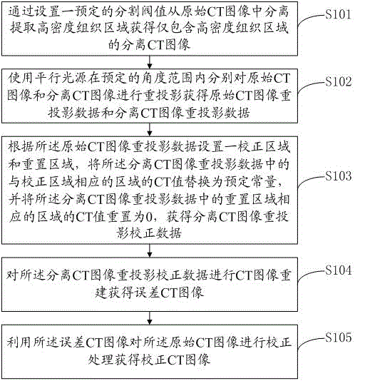
[0055] see figure 1 , figure 1 It is a flowchart of a preferred embodiment of the intraoperative CT image beam hardening artifact correction method of the present invention, as shown in the figure, including steps:

[0056] S101. Separating and extracting the high-density tissue region from the original CT image by setting a predetermined segmentation threshold to obtain a separated CT image containing only the high-density tissue region;

[0057] S102. Using a parallel light source to respectively reproject the original CT image and ...


PUM


Abstract

The invention discloses an intraoperative CT (Computed Tomography) image beam hardening artifact correction method and device. According to the method provided by the invention, a separated CT image only containing high-density tissues is separated from an original CT image, the two images are respectively processed by pre-projection, and then, the re-projection data are corrected, the corrected re-projection data are used for image reconstruction so as to obtain an error CT image, the original CT image is corrected through the error CT image, and finally, the corrected CT image is obtained. By the correction of the method provided by the invention, the artifact of the CT image is basically eliminated or weakened, and the aim of correction on the original image is realized.

Description

technical field [0001] The invention relates to the field of intraoperative CT image processing, in particular to a method and device for correcting intraoperative CT image beam hardening artifacts. Background technique [0002] Because intraoperative CT (a kind of CT medical diagnostic equipment) has the characteristics of small size, light weight, and flexible use, it is more and more widely used. Like traditional CT, intraoperative CT can also perform tomographic scanning on patients, which has attracted much interest. The slice image of the area can be used in early diagnosis and treatment, non-invasive diagnosis and other fields. Intraoperative CT has a wide range of applications and can be used in occasions that require CT scanning, such as operating rooms, ICU intensive care units, emergency rooms, etc. By using intraoperative CT, it can improve the success rate of operations and prolong the life of patients Purpose. [0003] However, a widespread artifact in the im...

Claims


Application Information

Patent Timeline
Patent Type & Authority Patents(China)
IPC IPC(8): G06T5/00G06T5/50A61B6/03
Inventor 薛国伟姜晓燕谢涛
Owner SHENZHEN ANKE HIGH TECH CO LTD
Why Eureka
Try Eureka