Method for segmenting cervix uteri liquid base cell image

A liquid-based cell and image segmentation technology, used in image analysis, image enhancement, image data processing, etc., can solve the problems of lack of cytoplasm, inability to segment cells and cell groups, and inability to segment adhering cell nuclei, etc. The effect of varying, reliable and precise segmentation results

Active Publication Date: 2012-12-19
SHENZHEN MICRO BIOLOGICAL TECH CO LTD
View PDF4 Cites 41 Cited by
  • Summary
  • Abstract
  • Description
  • Claims
  • Application Information

AI Technical Summary

Problems solved by technology

However, the active contour model used by Harandi et al. to extract the cytoplasmic boundary is sensitive to noise. When there are impurities, air bubbles, inflammatory cells and other objects in the background, the active contour will stop converging and the cytoplasmic boundary cannot be obtained.
The traditional method of segmenting the nucleus is usually to binarize the image directly in the cytoplasmic region. This region-based segmentation method will cause many false positive targets (such as cytoplasmic overlapping parts), and it is difficult to extract when the nuclei are stained unevenly. Accurate nuclei borders, additionally no s...

Method used


Image

  • Method for segmenting cervix uteri liquid base cell image
  • Method for segmenting cervix uteri liquid base cell image
  • Method for segmenting cervix uteri liquid base cell image

Examples


Embodiment Construction

[0021] The key technology of the present invention is to segment the cytoplasm and nucleus in the liquid-based cell image of the cervix. The technical principle of the present invention includes computer vision and image processing technology.

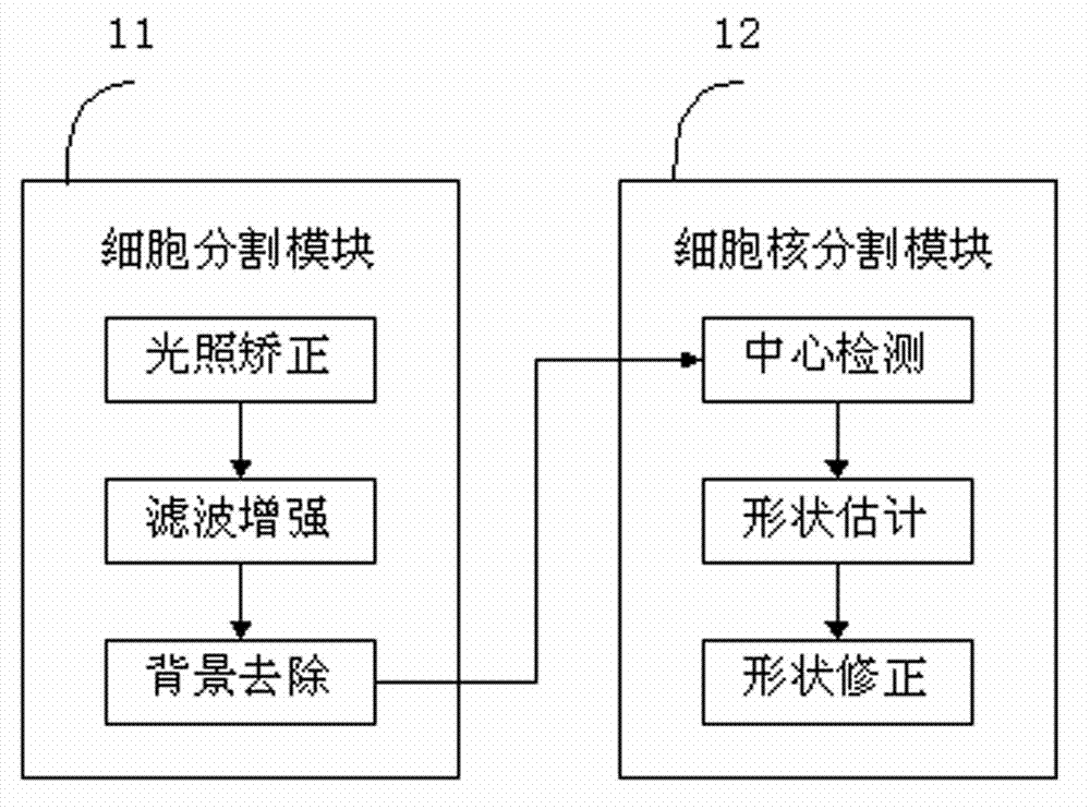
[0022] figure 1 It is a schematic diagram of a preferred embodiment of the present invention for cervical liquid-based cell image segmentation. The solution includes a cell segmentation module 11 and a cell nucleus segmentation module 12 . The cell segmentation module 11 is used to automatically exclude the background area to obtain the cytoplasm of single cells and cell groups, and the cell nucleus segmentation module 12 is used to automatically extract all the nucleus areas in the cytoplasm area, including cohesive nuclei.

[0023] The cervical liquid-based cell image segmentation method provided by the present invention comprises the following steps:

[0024] Step 1. Cell segmentation. First, light correction is performed on the...


PUM


Abstract

The invention discloses a method for segmenting a cervix uteri liquid base cell image. The method comprises the following steps of: 1, cell segmentation: performing illumination correction on an image, filtering noises and enhancing a cell boundary; and dividing the image into three types of cytoplasm, nucleus and background to eliminate a background class area; 2, nucleus segmentation: detecting centers of each of nucleuses in the cell area, estimating the approximate shapes of the nucleuses close to the centers of the nucleuses and correcting the shapes of the nucleuses to acquire an accurate boundary. The method is applied to cervix uteri cells and cell masses in a wide vision range, so that the phenomena of changed illumination, non-uniform dyeing and noise influence to a certain extent are avoided, and the cytoplasm, the single nucleus and adhesion nucleus can be segmented reliably and accurately.

Description

technical field [0001] The invention relates to cytopathology, computer vision and image processing methods, in particular to a method for automatic segmentation of cervical liquid-based cell images. Background technique [0002] Cervical cancer is one of the most threatening malignant tumors for women, and nearly 250,000 people die from cervical cancer every year in the world. In fact, cervical cancer is a curable cancer, as long as the cancer or HPV (human papillomavirus) infection can be detected early. At present, the most mature and effective method for clinical detection of early cervical cancer is liquid-based cytology combined with Pap staining. This preparation technique enables most cells to be spread evenly on the glass slide, thus facilitating the observation of cell nuclei and cytoplasm; this staining method enables the cells of different layers to display different colors, so that the cells of different layers can be differentiated to identify. It has been r...

Claims


Application Information

Patent Timeline
IPC IPC(8): G06T7/00G06T5/00
Inventor 唐盛张灵尹立东
Owner SHENZHEN MICRO BIOLOGICAL TECH CO LTD
Why Eureka
Try Eureka