

C-arm tomography imaging method using semi-accurate filtered back-projection
A filter back projection and tomographic imaging technology, applied in the field of biomedical imaging, can solve problems that affect the accuracy of reconstruction results, are not conducive to parallel processing of algorithms, and resampling of projection data, etc.
- Summary
- Abstract
- Description
- Claims
- Application Information
AI Technical Summary
Problems solved by technology
Method used
Image

Examples
Embodiment Construction
[0033] The present invention is specifically described below through the examples, it is necessary to point out that the present examples are only used to further illustrate the present invention, and can not be interpreted as limiting the protection scope of the invention, those skilled in the art can according to the above-mentioned present invention Some non-essential improvements and adjustments made in the content of the invention.
[0034] Embodiments of the present invention will be described in detail below with reference to the accompanying drawings.
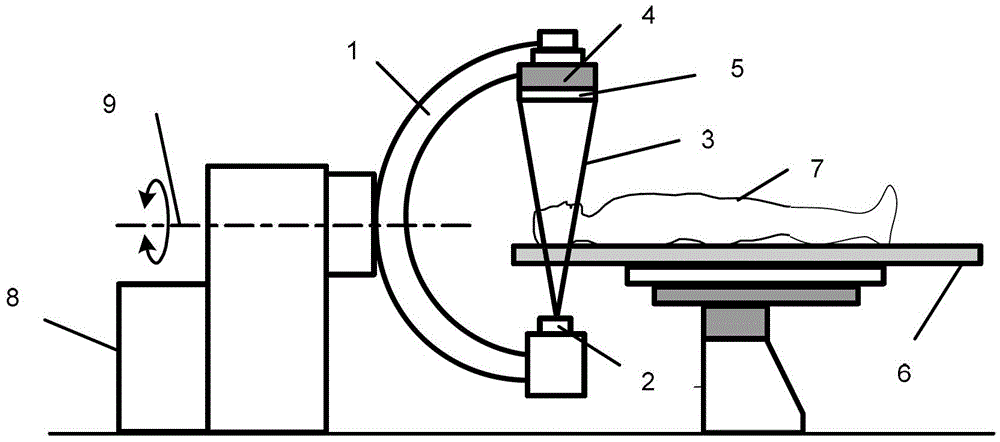
[0035] figure 1 Shown is a C-arm imaging device, which includes a type C-arm 1, one end of which is equipped with an X-ray source 2 for generating X-rays 3, which form a cone beam in space for emission; The other end of the arm 1 is equipped with a two-dimensional plane detector 4, the detector is composed of some array elements 5, and each array element 5 can sense the X-ray 3 attenuation signal passing through the pa...
PUM
 Abstract
Description
Claims
Application Information