P16 immune cell chemical reagent kit and polypeptide sequence in preparing same
An immunocytochemistry and kit technology, applied in the fields of peptides, peptide sources, biological tests, etc., can solve the problem of insufficient sensitivity of single cell detection
- Summary
- Abstract
- Description
- Claims
- Application Information
AI Technical Summary
Problems solved by technology
Method used
Image
Examples
Embodiment 1
[0047] Embodiment 1, kit assembly
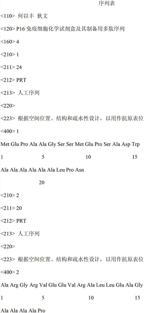
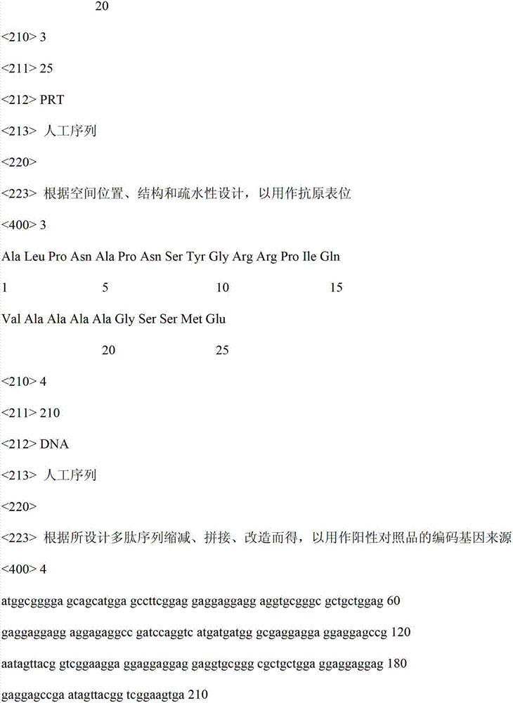
[0048] Three conjugates of keyhole limpet hemocyanin and polypeptides A, B and C (with glutaraldehyde as the coupling agent) were used to immunize mice respectively, and prepared according to the common monoclonal antibody preparation method against the above three polypeptide antigens Mouse anti-human P16 protein IgG monoclonal antibody production series (each production series contains 3 optimized and selected antibodies). Dilute the purified monoclonal antibody stock solution in phosphate buffer, prepare antibody mother solution at 1 mg / ml, and then mix any three antibody mother solutions at 1:1:1 (volume ratio) according to different combinations to ensure Each mixture combination contains and only contains three antibody members, and the three are from three different antibody preparation series. Then, using the obtained tri-antibody mixture (total antibody concentration: 1 mg / ml) and the three individual members of the mixture (ie: 1 ...
Embodiment 2
[0056] Embodiment two, the detection process of the sample to be tested
[0057] Take the 70% ethanol suspension containing women's cervical exfoliated cells, spread it on a glass slide (the coating area must be less than 5 square centimeters), and dry it at room temperature to become a cervical cell smear to be tested.
[0058] Take 2 microliters of component No. (1), add 98 microliters of component No. (6), dilute to become the primary antibody working solution, add dropwise to the cervical cell smear to be tested, covering all the cell coating area. Incubate for 1 hour at room temperature.
[0059] Use 10 ml of phosphate buffer solution (prepared by the experimenter, not provided in this kit) to wash away the above primary antibody working solution, and repeat three times.
[0060] Take 2 microliters (2) component, add 98 microliters (6) component, dilute to become the second antibody working solution, add dropwise to the cervical cell smear to be tested, covering the enti...
Embodiment 3
[0064] Embodiment three, the detection process of positive and negative reference substance
[0065] Cut open the plastic bags for positive and negative control substances, pour off all the 70% ethanol fixative solution, and gently rinse each petri dish three times with 10 ml of phosphate buffer solution (prepared by the experimenter, not provided in this kit).
[0066] Take 2 microliters of component (1), add 98 microliters of component (6), dilute to become the primary antibody working solution, and add dropwise to the petri dish of component (4) (ie: positive control substance) ; Also take 2 microliters of component (1), add 98 microliters of component (6), dilute to become the primary antibody working solution, and add dropwise to the culture of component (5) (ie: negative control substance) in the dish. Incubate for 1 hour at room temperature.
[0067] For each petri dish, wash away the above primary antibody working solution with 5 ml of phosphate buffer solution (prep...
PUM
 Abstract
Description
Claims
Application Information
 - R&D
- Intellectual Property
- Life Sciences
- Materials
- Tech Scout
- Unparalleled Data Quality
- Higher Quality Content
- 60% Fewer Hallucinations
Browse by: Latest US Patents, China's latest patents, Technical Efficacy Thesaurus, Application Domain, Technology Topic, Popular Technical Reports.
© 2025 PatSnap. All rights reserved.Legal|Privacy policy|Modern Slavery Act Transparency Statement|Sitemap|About US| Contact US: help@patsnap.com


