Method and relevant kit for detecting ALK gene status based on rare cells

A rare cell and kit technology, applied in the field of medical diagnostics, can solve the problems of lack of comprehensive and holistic evaluation of histological detection, inability to provide real-time monitoring information, and dynamic changes in molecular information.

Inactive Publication Date: 2015-11-25
北京莱尔生物医药科技有限公司
View PDF5 Cites 0 Cited by
  • Summary
  • Abstract
  • Description
  • Claims
  • Application Information

AI Technical Summary

Problems solved by technology

[0011] 1. As an invasive histological test limited by the location of the lesion, there are limitations such as difficulty or inability to obtain specimens, and repeated postoperative specimens cannot be obtained, resulting in the inability of tissue specimens to provide real-time monitoring information
[0012] 2. And affected by treatment, etc., the molecular information of tumor cells may change dynamically
For example, some studies have reported that breast cancer patients who received neoadjuvant

Method used


Image

  • Method and relevant kit for detecting ALK gene status based on rare cells
  • Method and relevant kit for detecting ALK gene status based on rare cells
  • Method and relevant kit for detecting ALK gene status based on rare cells

Examples


Embodiment 1

[0123] Testing a pair of blood samples using the kit

[0124] As shown in steps 1-6 of the above kit use method, test blood samples from 30 normal people, 40 benign lung diseases, and 40 non-small cell lung cancer patients. The method is to add 3.2mL blood samples to a 50mL centrifuge tube, Add CS1 working solution, centrifuge at 650×g for 5 minutes, aspirate and discard the supernatant; add CS2 working solution for lysis, centrifuge at 650×g for 5 minutes, aspirate and discard the supernatant; add a certain amount of CS1 working solution again, add magnetic particles to mix Suspension 200uL, shake well for 20 minutes; absorb all the mixed liquid, superimpose it on the CS3 separation medium, centrifuge at 300×g for 5 minutes; absorb the liquid except the magnetic particle precipitation into a 15mL centrifuge tube, add CS1 working solution to 15mL , centrifuge at 950×g for 5 minutes, discard the supernatant; add 1mL CS1 working solution, mix by pipetting and add to a new 2mL ce...

Embodiment 2

[0127] The preparation of kit of the present invention
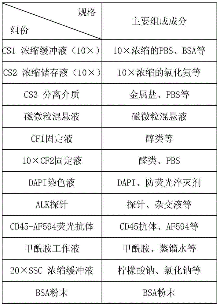
[0128] Kit one:

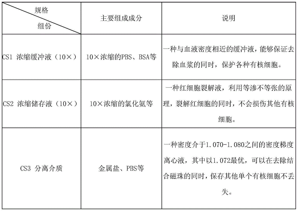
[0129] CS1 Concentrated Buffer (10×):

[0130] Each 1000mL water contains 60gBSA, 5 packs of PBS powder (2L / packet), 100mL0.5M EDTA, 0.8mLProclin300.

[0131] CS2 concentrated stock solution (10×):

[0132] Weigh 82.9gNH per 1000mL of water 4 Cl, 10gKHCO 3 , 0.37gEDTA, water and 0.8mLProclin300, fully stirred and dissolved, constant volume, and prepared as a 10X concentrated solution.

[0133] CS3 separation medium:

[0134] Dilute the gradient centrifugate with a density of 1.077, and test the density during the dilution process to make the density between 1.070-1.075.

[0135] Magnetic particle suspension:

[0136] Adjust the concentration of CD45 antibody to 1mg / mL, and incubate with streptavidin immunomagnetic beads at a ratio of 100uL:1mL for 1h to prepare a suspension of magnetic particles.

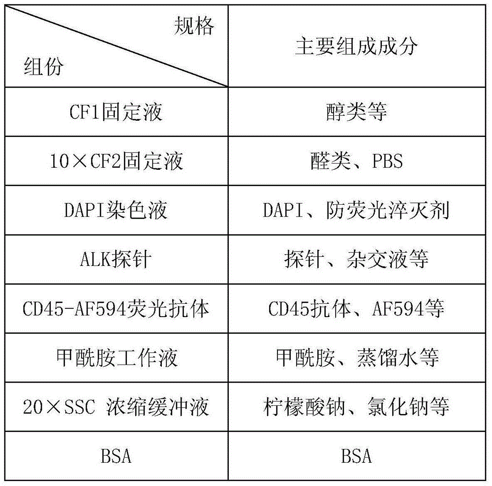
[0137] CF1 fixative:

[0138] Mix PEG and absolute ethanol so that the final concentration of PEG...

Embodiment 3

[0155] Use the test kit of the present invention to detect a pair of other body fluid samples

[0156] Note: This kit is not only suitable for blood, but also for the detection of rare cells in other body fluids, such as pleural effusion, ascites, toilet fluid, amniotic fluid, etc., but not limited to these types of body fluids.

[0157] Using this kit to detect 6 cases of lung cancer pleural effusion, 1 case found ALK rearrangement positive cells, the number was 4.


PUM


Abstract

The invention relates to a method and a relevant kit for detecting ALK gene status based on rare cells. The method comprises the following steps: step 1, taking a blood sample or a body fluid sample, wherein the sample contains a mixed cell population comprising circulating tumor cells (CTCs) or other rare cells, and white blood cells; step 2, diluting the sample by adopting a buffer solution, so as to remove plasma and recover the CTCs or the other rare cells; step 3, splitting by adopting red blood cell splitting solution, so as to remove red blood cells and recover the CTCs or the other rare cells; step 4, uniformly mixing magnetic particles coated by relevant human-leukocyte-resisting antigen-antibodies and the recovered cells obtained in the step 3 for incubation, and mixing to form a magnetic particle-white blood cell mixture; step 5, separating the magnetic particle-white blood cell mixture from other cells through centrifugation, and thus obtaining the mixed cell population containing CTCs or other rare cells, and small quantity of white blood cells; and step 6, determining the ALK gene rearrangement state of CTCs or other rare cells by adopting a method of combining the immunofluorescence cytochemistry and fluorescence in situ hybridization.

Description

technical field [0001] The present invention relates to medical diagnostics, in particular to the detection of tumor cells. More specifically, the present invention relates to diagnostic methods for detecting cancer and evaluating treatment options for patients. Background technique [0002] Circulating tumor cells (Circulating Tumor Cell, CTC) refer to the tumor cells that enter the blood circulation, which can fall off into the blood from the primary tumor or metastases, and may also enter the blood before forming solid tumor lesions. Tumor cells invade the circulatory system, and most of them die within a short period of time due to the body’s immune recognition, mechanical killing, and self-apoptosis, and only a few survive and plant in distant or primary tissues and organs, and further develop into metastases. The most direct factor leading to tumor metastasis and recurrence. [0003] CTC is a "liquid biopsy" sample that can represent the primary tumor. Peripheral blo...

Claims


Application Information

Patent Timeline
IPC IPC(8): C12Q1/68G01N33/573G01N33/58
CPCC12Q1/6841C12Q1/6886C12Q2600/106C12Q2600/156G01N33/573G01N33/582C12Q2543/10
Inventor 詹厅杨瑛杰马妤婕
Owner 北京莱尔生物医药科技有限公司
Why Eureka
Try Eureka