
Preparation and application of a monoclonal antibody against alveolar coccoid cyst wall tissue (germinal layer, corneum cortex)

A technology of monoclonal antibody and corneum, which is applied in the field of biotechnology and cell engineering, can solve the problems of lack of monoclonal antibody and inability to develop

Active Publication Date: 2018-10-19
王昕 +2
View PDF0 Cites 0 Cited by
  • Summary
  • Abstract
  • Description
  • Claims
  • Application Information

AI Technical Summary

Problems solved by technology

However, there has been a lack of monoclonal antibodies with strong specificity and sensitivity required for the use of immunohistochemical methods, and it is impossible to carry out

Method used


Image

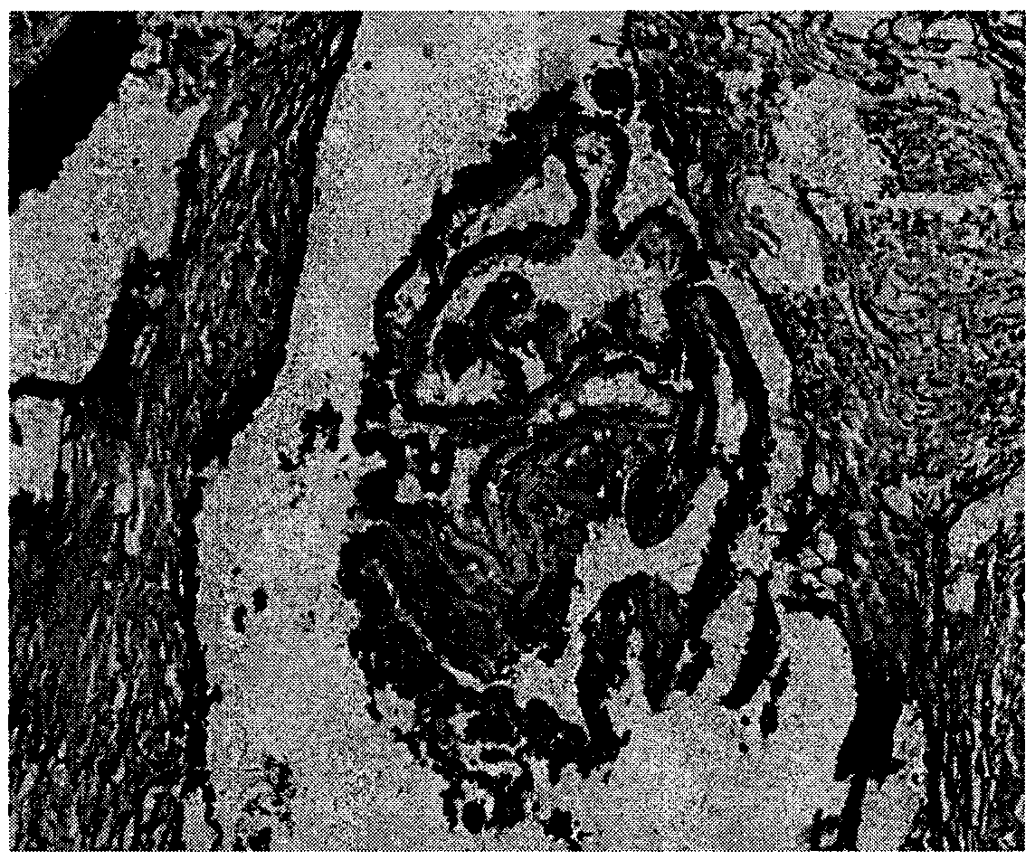
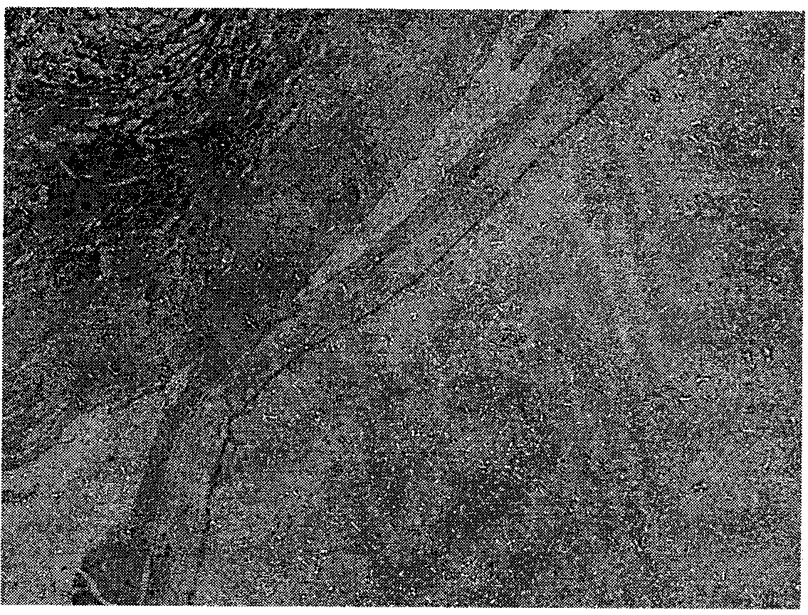
  • Preparation and application of a monoclonal antibody against alveolar coccoid cyst wall tissue (germinal layer, corneum cortex)
  • Preparation and application of a monoclonal antibody against alveolar coccoid cyst wall tissue (germinal layer, corneum cortex)
  • Preparation and application of a monoclonal antibody against alveolar coccoid cyst wall tissue (germinal layer, corneum cortex)

Examples


Embodiment 1

[0016] Example 1 - Establishment of cell lines

[0017] 1. Antigen Preparation

[0018] Materials: gerbils infected by alveolar coccus, petri dishes, surgical instruments, 50ml round-bottom centrifuge tubes for disinfection, 0.9% sodium chloride injection, tissue homogenizer (Ningbo Xinzhi XHF-D), cell ultrasonic pulverizer ( Ningbo Xinzhi JY92-II).

[0019] Procedure: The gerbils infected with alveolar coccidia were killed by over-anesthesia with ether. Soak in 75% ethanol for 15 minutes to disinfect the body surface. Open the abdomen according to the anatomical level. Cut well-grown alveolar coccidioid tissue in the abdominal cavity and put it into a petri dish, add 6ml of 0.9% sodium chloride injection. Carefully remove host tissue and blood vessels from the surface of the worm with ophthalmic scissors. Take 15g of processed hydatid tissue and put it into a 50ml centrifuge tube.

[0020] Break up the tissue with a tissue homogenizer at 2800 rpm for 3 minutes. Centrif...

Embodiment 2

[0049] Embodiment 2-expansion culture and cryopreservation of cell lines

[0050] 1. Expansion of cell lines

[0051] The obtained positive cells were taken out from the 96-well plate and put into a 24-well plate for expanded culture. The culture medium is complete culture medium.

[0052] 2. Cryopreservation of hybridoma cells

[0053] Materials: Cell cryopreservation medium: 25ml of calf serum, 20ml of RPMI1640 incomplete culture medium, 5ml of dimethyl sulfoxide were mixed as cell cryopreservation medium. -70°C refrigerator with liquid nitrogen, cryopreservation tubes.

[0054] Steps: observing the monoclonal hybridoma cells expanded and cultured in the 24-well culture plate, when the bottom of the well is covered, it is observed that the cells grow well, and the living cells are greater than 90%. Blow off the cells at the bottom of the well with complete cell culture medium. 2-3 wells were collected into centrifuge tubes. After centrifugation at 2000rpm for 5min, the...

Embodiment 3

[0055] Example 3-Mass preparation of monoclonal antibody (preparation of ascites)

[0056] Materials: 6-8 weeks old Balb / c mice, liquid paraffin, cell counting plate, RPMI1640 incomplete culture medium.

[0057] Steps: mouse sensitization: Balb / c mice were sensitized with liquid paraffin, and the injection volume was 500ul / mouse. Ascitic fluid was prepared 10 days later. Injection of cells: Collect hybridoma cells and wash the cells twice with RPMI1640 incomplete culture medium. Take 1 to 1.5 million cells and inject them into the abdominal cavity of mice. After one week, it can be seen that the mice are inactive and the abdominal cavity of the mice is enlarged. Ascites collection: 7 to 10 days after cell inoculation, ascites can be seen. Closely observe the health status of the animals and signs of ascites. When ascites is as much as possible and the mice are on the verge of death, kill the mice and inhale the ascites into the test tube. The collected ascites was stored in ...


PUM


Abstract

The invention discloses preparation and applications of echinococcus multilocularis resistant vesicle wall tissue (malpighian layer and cuticle layer) monoclonal antibody. The larva of echinococcus multilocularis parasitizes in human body and causes multilocular hydatidosis which is also known as echinococcus multilocularis disease. The larva usually parasitizes in the liver, and the disease is usually confused with tumors or other diseases. The invention relates to the scientific research, diagnosis, and treatment of echinococcus multilocularis disease, and provides a mouse-originated hybridoma cell strain. The monoclonal antibody secreted by the mouse-originated hybridoma cell strain can carry out specific binding with vesicle wall tissue (malpighian layer and cuticle layer) of echinococcus multilocularis, so the antibody can be used to carry out immunohistochemical specific staining on pathological tissues of the echinococcus multilocularis disease. The antibody titer can reach 1:105, and the antibody has a very strong sensitivity and specificity. The antibody can be used for the immunological pathological diagnosis of the echinococcus multilocularis disease. The monoclonal antibody can also be coupled with chemotherapy drugs or radionuclide to prepare target drugs and diagnosis reagents. The antibody can be applied to the scientific research related with echinococcus multilocularis too.

Description

technical field [0001] The invention belongs to the fields of biotechnology and cell engineering. It relates to a hybridoma cell line secreting monoclonal antibody against alveolar cyst wall tissue and the antibody secreted therefrom, and its application in the diagnosis and treatment of multilocular echinococcosis (alveolar coccosis). Background technique [0002] Multilocular hydatid disease (Multilocular hydatid disease), also known as alveolar coccidiosis, is a parasitic disease caused by alveolar coccidia, the larvae of Echinococcus multilocularis. Multilocular echinococcosis is mainly prevalent in pastoral and semi-agricultural and semi-pastoral areas in western China, especially in Xinjiang, Qinghai, Gansu, Ningxia, Tibet, Inner Mongolia and western Sichuan. It caused great losses to the local economy. The adult worms of Echinococcus multilocularis live in the small intestines of final hosts such as foxes and dogs. When rodents and humans swallow the eggs excreted w...

Claims


Application Information

Patent Timeline
Patent Type & Authority Patents(China)
IPC IPC(8): C12N5/20C07K16/18G01N33/577A61K39/395A61P33/10C12R1/91
Inventor 王昕陈建平刘巧凤
Owner 王昕
Learn More