A method for segmenting renal vascular compartments based on medical images

A technology of medical imaging and kidney, applied in the field of medical image analysis

Inactive Publication Date: 2017-05-10
NORTHEASTERN UNIV LIAONING +1
View PDF4 Cites 0 Cited by
  • Summary
  • Abstract
  • Description
  • Claims
  • Application Information

AI Technical Summary

Problems solved by technology

The shortcoming of this method lies in the selection of the scanning phase. After the contrast agent is injected, the kidney will be scanned in multiple phases, and under different scanning phases, the areas with bright signals in the kidney will have various changes. Therefore, scanning images of different phases will produce different corticomedullary segmentation results.

Method used


Image

  • A method for segmenting renal vascular compartments based on medical images
  • A method for segmenting renal vascular compartments based on medical images
  • A method for segmenting renal vascular compartments based on medical images

Examples


Embodiment Construction

[0046] The specific implementation manners of the present invention will be described in detail below in conjunction with the accompanying drawings.

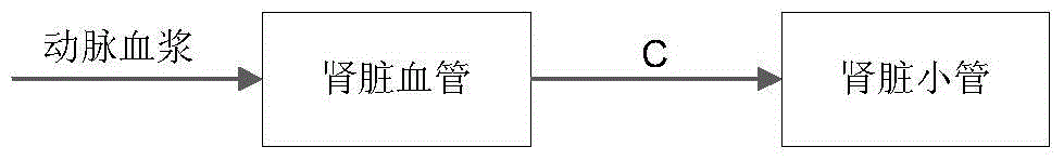
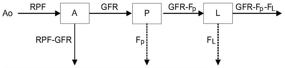
[0047] A method for segmenting renal vascular compartments based on medical images, such as Figure 14 shown, including the following steps:

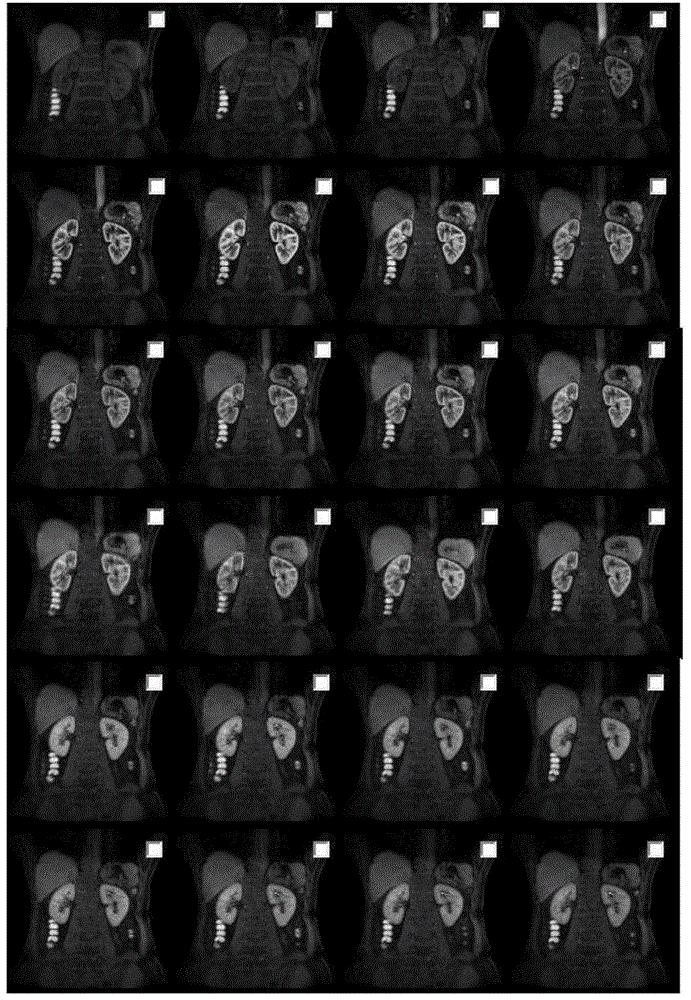
[0048] Step 1: Obtain all the images of the dynamic contrast-enhanced scan of the kidney;

[0049] Step 1-1: Obtain a phased kidney scan image without injecting contrast agent;

[0050] Step 1-2: Inject contrast agent, and perform an enhanced kidney scan every 3 seconds;

[0051] Steps 1-3: Continuously acquire kidney enhanced scan images of multiple phases to obtain all images of kidney dynamic enhanced scan;

[0052] The image scanning was all completed on the GE 3.0T HDx machine, using the body phased array coil. After the positioning scan, the conventional sequence scan was performed first, and then the MRU and phase contrast MRA were performed. Scanning parameters TR = 4. 7 ms, T...


PUM


Abstract

The invention provides a method for segmenting kidney blood vessel atrioventricular on basis of medical images. The method includes that all images of kidney dynamic enhancement scan are obtained; kidney enhancement scan image data of different scan period phases are subjected to registration; a kidney area is obtained by segmentation from kidney enhancement scan images; a TDC curve of each voxel in kidney area images is drawn; time TTP of reaching peak concentration in the TDC curve of each voxel in the kidney area images is calculated; a TTP feature map of kidney is generated; the TTP feature map of the kidney is subjected to thresholding by a self-adaption threshold value method; a kidney blood vessel atrioventricular segmentation result is obtained. According to the method for segmenting kidney blood vessel atrioventricular on basis of the medical images, time feature images about kidney contrast agent concentration are generated on basis of dynamic enhancement kidney scan images, and blood vessel atrioventricular segmentation extraction can be achieved by an image processing method on basis of the time feature images.

Description

technical field [0001] The invention belongs to the technical field of medical image analysis, and in particular relates to a method for segmenting renal blood vessel compartments based on medical images. Background technique [0002] Analyzing the filtering function of the kidney based on medical images will become a development trend in clinical applications in the future, and a large number of related studies have been carried out so far. In this analysis process, the compartment model is a widely used analysis model. In the study, the compartmental model of the kidney is divided into different models, including two-compartment model and three-compartment model, which are all used to describe the filtration process of blood in the whole kidney. The same thing is that no matter the several-compartment model, it will include a division of the vascular compartment, that is, the blood is filtered from the vascular compartment to other compartments. In the process of calcula...

Claims


Application Information

Patent Timeline
Patent Type & Authority Patents(China)
IPC IPC(8): G06T7/00A61B6/03
Inventor 李宏孙浩然康雁金时开徐谢平鲍楠孙航田实
Owner NORTHEASTERN UNIV LIAONING
Why Eureka
Try Eureka