Eureka AIR delivers breakthrough ideas for toughest innovation challenges, trusted by R&D personnel around the world.

Assessment of vascular compartment volume for PET modelling

a vascular compartment and pet technology, applied in the field of ##positron emission tomography (pet), can solve the problems of inability to accurately estimate vsub>b /sub>from the dynamic pet image with pkm, method does not take into account variation which may arise from patient physiology and anatomy, and achieves accurate localization of metabolic activity

Active Publication Date: 2011-12-13
SIEMENS MEDICAL SOLUTIONS USA INC
View PDF7 Cites 4 Cited by
  • Summary
  • Abstract
  • Description
  • Claims
  • Application Information

AI Technical Summary

Benefits of technology

The patent text describes a technique called Positron Emission Tomography (PET) that uses a radioactive tracer to create a three-dimensional map of functional processes in the body. The technique involves injecting the tracer into the body and detecting the gamma rays emitted as the tracer decays. The resulting images show how the tracer is distributed and how it accumulates in different tissues over time. The technique can provide important information about the behavior of the body in real-time, but there are limitations in accurately estimating the concentration of the tracer in certain tissues. The patent text also describes the use of Magnetic Resonance Imaging (MRI) to estimate the concentration of blood vessels in the brain. The text highlights the limitations of each technique and proposes a method to combine the two to improve the accuracy of estimating the ratio of blood vessel volume.

Problems solved by technology

Estimation of VB from the dynamic PET image with PKM is possible, but may not be very accurate, particularly since the useful time frames of the dynamic images for estimation of VB are the earlier ones, when the tracer is still predominantly in the blood and starts to be rapidly taken up in the cell.
However, because it is a rapid process, the early time frames of PET cover a short time interval and are therefore very noisy, making the estimation of VB relatively unreliable.
However, this method does not take into account variation which may arise from the patient physiology and anatomy as well as pathophysiology of the disease (for example, in cancerous tumours, angiogenesis involves the local creation of a massive amount of blood vessels, which increases VB significantly).
The technique is used routinely in studies, but the estimation of VB lacks robustness, which impacts the estimation of the rate constants, which is what the clinician is really after.
Unfortunately, there are a number of limitations which prevent the use of contrast enhanced MRI to model cellular processes as finely as PET can, mainly because a) the contrast agents need to be injected in much higher concentrations and b) the agents are usually much larger molecules which do not penetrate easily into the cell.

Method used


Image

  • Assessment of vascular compartment volume for PET modelling
  • Assessment of vascular compartment volume for PET modelling

Examples


Embodiment Construction

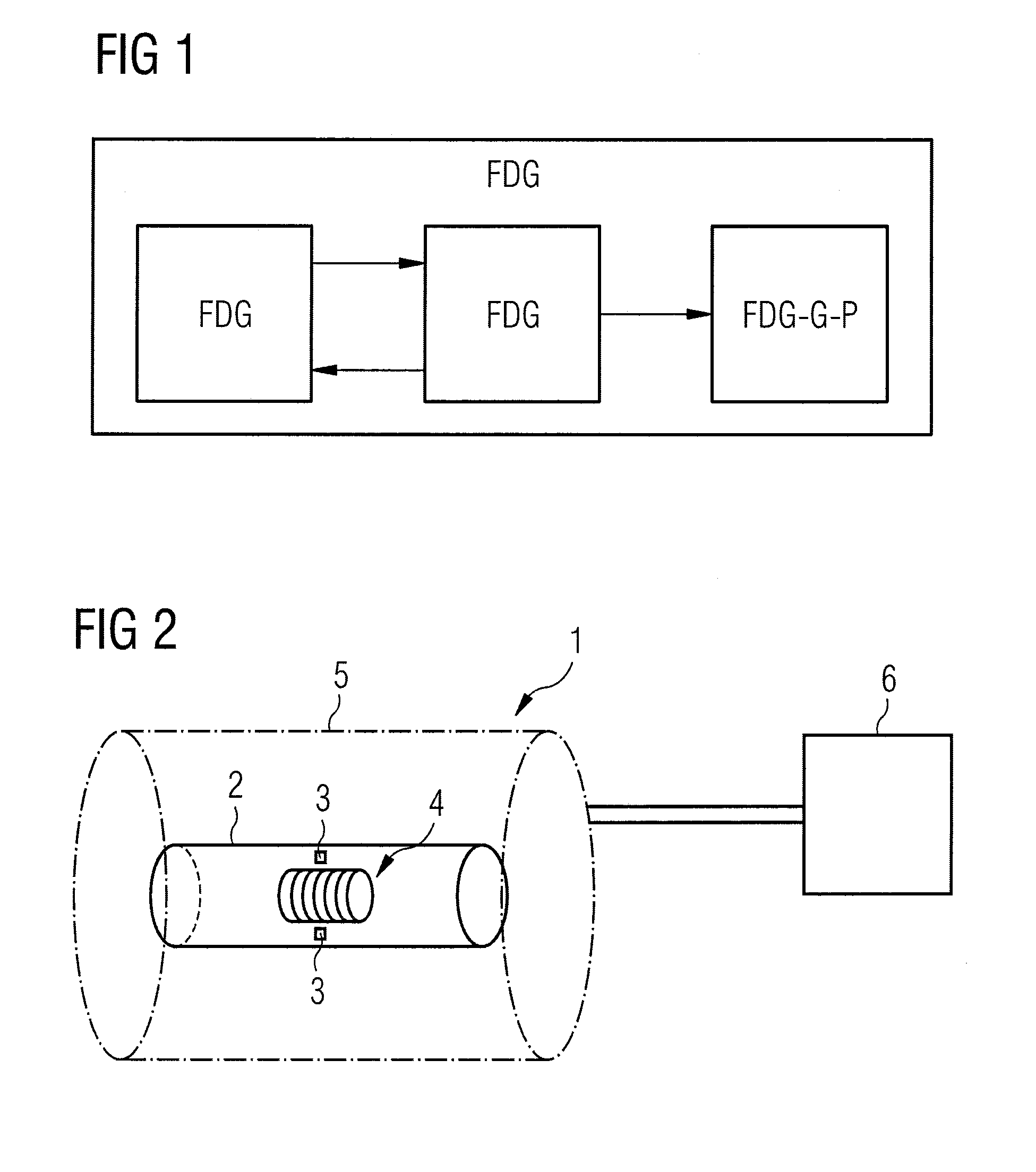
[0020]Referring to FIG. 2, apparatus of the invention, generally designated 1, includes a PET scanner 2 having a ring of scintilators 3 and a series of RF coils 4 located in the main magnet 5 of an MRI scanner (other components not shown).

[0021]Such a combination of PET and MRI scanning apparatus enables simultaneous and iso-volumic acquisition of a volume of interest in PET and MRI. With such a device, a combination of a radioactive PET compound and a blood stream MRI contrast agent is injected simultaneously, and both PET and MR dynamic images are acquired.

[0022]The PET / MRI apparatus are controlled by processor 6. Software applications allow user interaction to set parameters for a particular protocol and initiate scanning. Data acquisition and storage would also typically be controlled by processor 6.

[0023]The dynamic MRI is acquired over the early part of the imaging protocol as the blood stream information quickly reaches some equilibrium, and it is possible to estimate VB from...


PUM


Abstract

A method is described for acquiring and analysing the data produced by Positron Emission Tomography (PET), which method provides for an accurate estimation of vascular compartment volume, VB. An MRI scan and the PET scan is performed simultaneously and the results of the former is used to derive a value for VB. The value so derived is then used in pharmacokinetic modelling along with the results of the functional scan.

Description

[0001]The invention is concerned with the estimation of vascular compartment volume for pharmo-kinetic modelling of Positron Emission Tomography (PET) data.BACKGROUND AND SUMMARY OF THE INVENTION[0002]Positron Emission Tomography (PET) is a nuclear medical imaging technique which provides a three-dimensional map of functional processes in the human or animal body.[0003]A radioactive (positron emitting) tracer isotope is incorporated in a metabolically active molecule such as 18-F fluoro-2-deoxy-2-glucose (FDG, a sugar analogue) which is ingested in a subject. Other radiolabelled molecules which may be used in PET include 3′-deoxy-3′-[18-F]-fluorothymidine (FLT) and 6-[(18)F]fluoro-L-DOPA (FDOPA). These molecules can be imaged by a scanner or camera which detects and records the gamma type radiation resulting from a collision between an emitted positron and an electron in the surrounding matter.[0004]The radiation so produced is released as two photons travelling in near opposite dir...

Claims


Application Information

Patent Timeline
Patent Type & Authority Patents(United States)
IPC IPC(8): A61B5/05
CPCA61B5/055A61B6/037A61B6/5247G01R33/481G01R33/281G01R33/48G01T1/2978G01T1/2985
Inventor DECLERCK, JEROMEHAKL, KEVIN SCOTT
Owner SIEMENS MEDICAL SOLUTIONS USA INC
Why Eureka
Eureka Blog
Learn More