System and method to analyze blood parameters using magnetic resonance imaging

a magnetic resonance imaging and blood parameter technology, applied in the field of magnetic resonance imaging system and methods, can solve the problems of inability to produce voxel-by-voxel oef maps, inability to cleanly target blood from post-capillary veins, and inability to accurately measure sao2

Inactive Publication Date: 2015-02-17
THE GENERAL HOSPITAL CORP
View PDF4 Cites 13 Cited by
  • Summary
  • Abstract
  • Description
  • Claims
  • Application Information

AI Technical Summary

Benefits of technology

[0014]The present invention overcomes the aforementioned drawbacks by providing a system and method for accurately measuring SaO2 directly from compartmentally-isolated blood using magnetic resonance imaging. Furthermore, the present invention is able to generate accurate OEF and CMRO2 maps from the measurements, since SaO2 is an intrinsic property of blood alone. Further still, the present invention is able to generate kinetic curves of oxygen exchange, thus, giving detailed insight into oxygen exchange dynamics.
[0015]One aspect of the invention provides an ability to isolate blood signal based on velocity range and acceleration. Velocity and acceleration are properties of blood that are dependent on the containing vascular compartment. A velocity range defines a specific population of blood; however, for all but capillary blood, a velocity range selects an arterial pool of blood and the analogous venous pool. The present invention allows discrimination between the two based on the fact that arterial blood decelerates as it approaches the capillaries, while venous blood accelerates while approaching larger veins. Thus, the combination of a delay (inflow time) framed by velocity selection modules, allows targeting of blood that either decelerates (i.e. arterial blood) or accelerates (i.e. venous blood). In this way, it is possible to specifically target arterial / arteriolar or venous / venular blood compartments, despite similar velocity range. Additionally, since stationary spins are eliminated by control-tag subtraction, signal is clearly attributed to spins originating in blood. Because of the precision targeting of arteriolar and venular blood signal, OEF (and subsequently CMRO2) can be measured on a voxel-by-voxel basis.

Problems solved by technology

Previous MR methods exist to measure OEF, but are limited.
The major challenge with such methods has been to separate the MR signal from various arterial, capillary, and venous compartments, whose blood will have different oxygen concentrations.
For example, these methods are unable to cleanly target blood from post-capillary venules and cannot produce OEF maps on voxel-by-voxel basis.
Susceptibility methods are particularly limited as they require manual, visual identification of draining veins, as identified by a functional activation experiment.
These methods also require precise knowledge of vessel geometry and cannot be used to generate absolute Yv or OEF maps.
Moreover, the static-dephasing-regime theory used may not hold for capillaries.
However, these approaches are hindered by restrictive assumptions, are confounded by signal arising from tissue, and / or are unable to produce accurate OEF maps on a voxel-by-voxel basis.

Method used


Image

  • System and method to analyze blood parameters using magnetic resonance imaging
  • System and method to analyze blood parameters using magnetic resonance imaging
  • System and method to analyze blood parameters using magnetic resonance imaging

Examples


Embodiment Construction

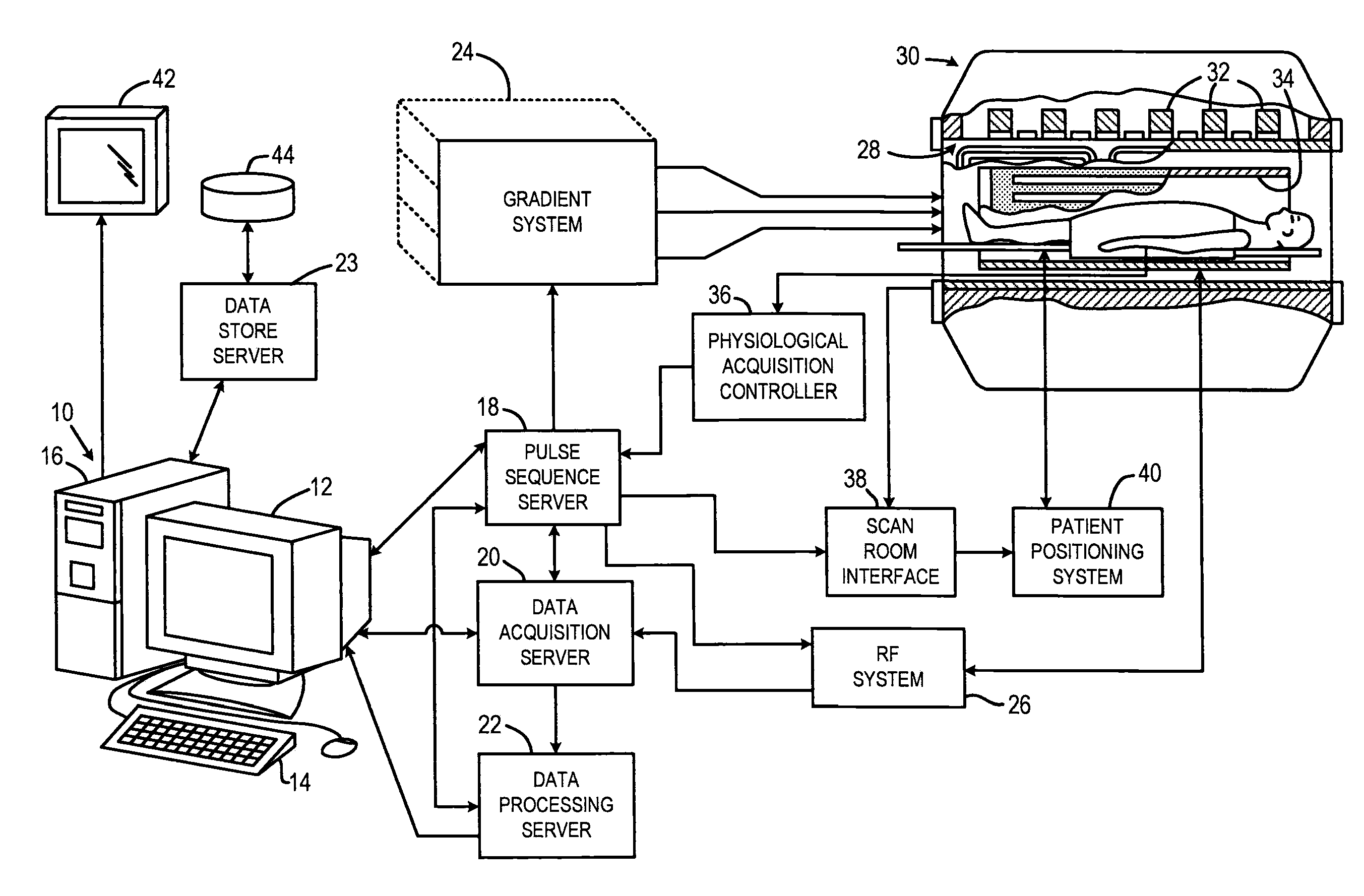
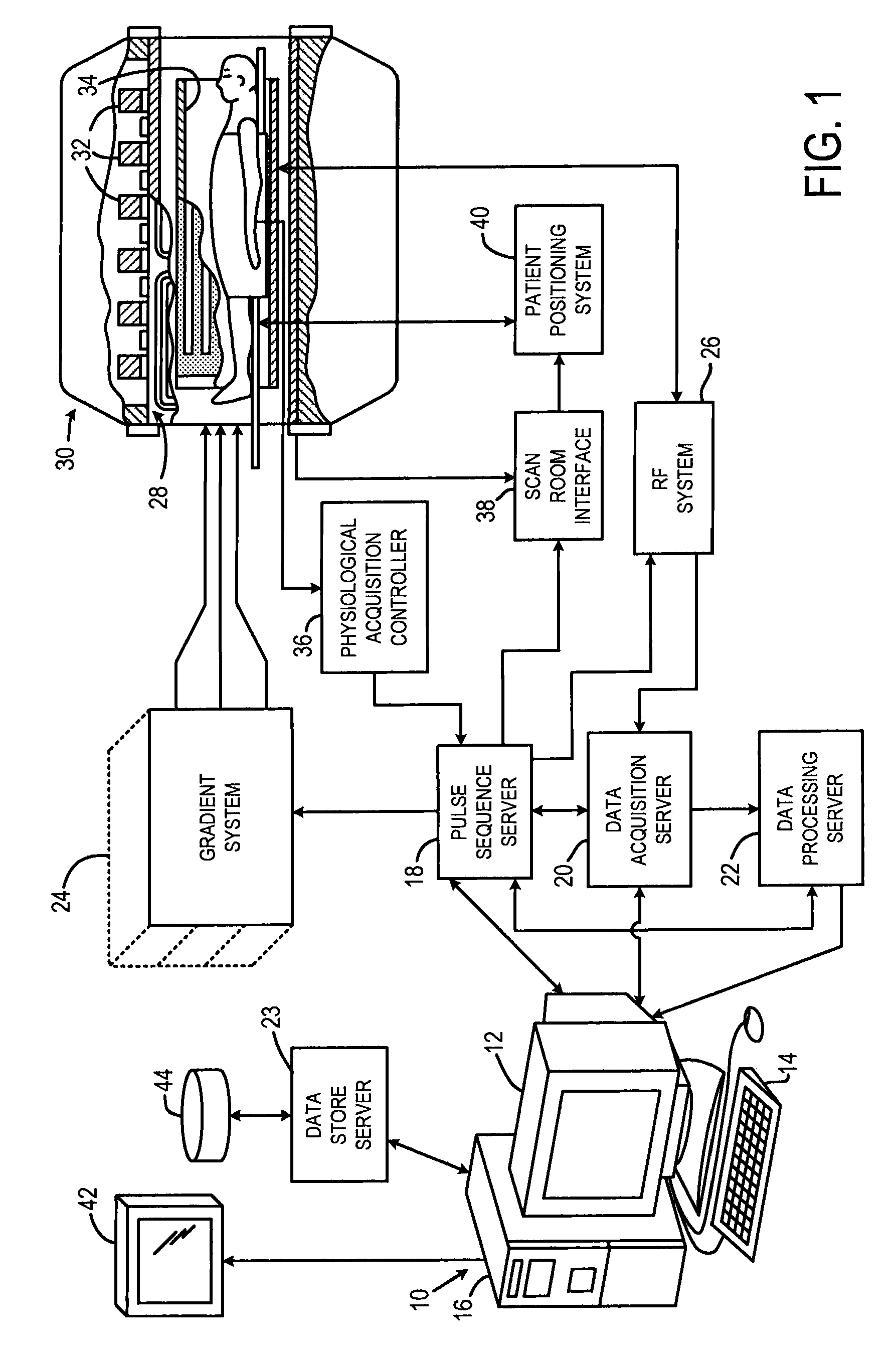
[0027]Referring particularly to FIG. 1, the present invention is employed using an MRI system. The MRI system includes a workstation 10 having a display 12 and a keyboard 14. The workstation 10 includes a processor 16 that is a commercially available programmable machine running a commercially available operating system. The workstation 10 provides the operator interface that enables scan prescriptions to be entered into the MRI system. The workstation 10 is coupled to four servers: a pulse sequence server 18; a data acquisition server 20; a data processing server 22, and a data store server 23. The workstation 10 and each server 18, 20, 22 and 23 are connected to communicate with each other.

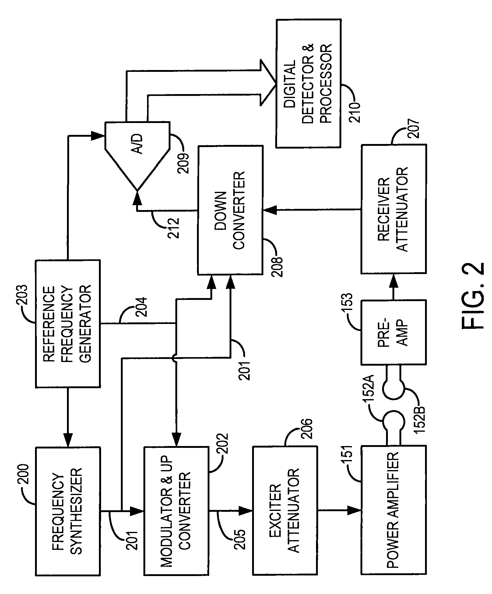
[0028]The pulse sequence server 18 functions in response to instructions downloaded from the workstation 10 to operate a gradient system 24 and an RF system 26. Gradient waveforms necessary to perform the prescribed scan are produced and applied to the gradient system 24 that excites gradient co...


PUM


Abstract

A system and method for accurately producing MR images of selected vascular compartments includes employing a control scan and a tag scan, each including velocity selective modules that suppress signal from blood flowing faster than a given cutoff velocity, to acquire control and tag sets of NMR data that may be subtracted to produce a compartment-specific MR image that is substantially free of information from stationary tissues and blood outside the selective vascular compartments. Accordingly, physiological parameters, such as oxygen saturation (SaO2), oxygen extraction fraction (OEF), and cerebral metabolic rate of oxygen (CMRO2), can be generated from the compartment-specific images. Further still, kinetic curves of oxygen exchange can be created, thus providing detailed insight into oxygen exchange dynamics.

Description

CROSS REFERENCE TO RELATED APPLICATIONS[0001]This application claims the benefit of U.S. Provisional Application 61 / 041,037, filed Mar. 31, 2008, which is incorporated herein in its entirety for all purposes.STATEMENT REGARDING FEDERALLY SPONSORED RESEARCH[0002]This invention was made with government support under 1R01EB007942-01A2 awarded by the National Institutes of Health. The government has certain rights in the invention.FIELD OF THE INVENTION[0003]The present invention relates to a system and methods for magnetic resonance imaging (MRI). More particularly, the present invention relates to targeting MRI signal from blood in specific vascular compartments using velocity selective pulses, accurately measuring oxygen saturation (SaO2) from compartmentally-isolated MR signal, generating accurate oxygen extraction fraction (OEF) and cerebral metabolic rate of oxygen (CMRO2) maps from the measurements, and generating kinetic curves of oxygen exchange to provide detailed insight into...

Claims


Application Information

Patent Timeline
Patent Type & Authority Patents(United States)
IPC IPC(8): A61B5/05G01V3/00G06K9/00A61B5/055A61B5/145G01R33/56G01R33/563
CPCA61B5/055G01R33/5607G01R33/56366A61B5/145G01R33/56316
Inventor BOLAR, DIVYA S.ADALSTEINSSON, ELFARROSEN, BRUCE R.SORENSEN, A. GREGORY
Owner THE GENERAL HOSPITAL CORP
Why Eureka
Try Eureka