Method for rapidly and accurately focusing and scanning pathological section tissue on basis of image collection device

An image acquisition device and pathological slice technology, applied in the field of scanning, can solve problems such as inaccuracy and slow focusing speed, and achieve the effect of improving scanning speed and image quality

Active Publication Date: 2015-03-04
KONFOONG BIOTECH INT
View PDF6 Cites 22 Cited by
  • Summary
  • Abstract
  • Description
  • Claims
  • Application Information

AI Technical Summary

Problems solved by technology

[0008] In order to improve the scanning speed and scanning quality more effectively on the basis of the prior art, the present invention provides a method for fast and accurate focusing scanning of pathological slice tissue based on an image acquisition device. This method obtains a rough virtual plane, and then continuously correct the movement track of the Z-axis according to the image data during the scanning process, thus well solving a series of problems such as slow focusing speed and inaccuracy

Method used


Image

  • Method for rapidly and accurately focusing and scanning pathological section tissue on basis of image collection device
  • Method for rapidly and accurately focusing and scanning pathological section tissue on basis of image collection device
  • Method for rapidly and accurately focusing and scanning pathological section tissue on basis of image collection device

Examples


Embodiment 1

[0043] (1) A method for fast and accurate focus scanning of pathological section tissue based on an image acquisition device, the steps of which include:

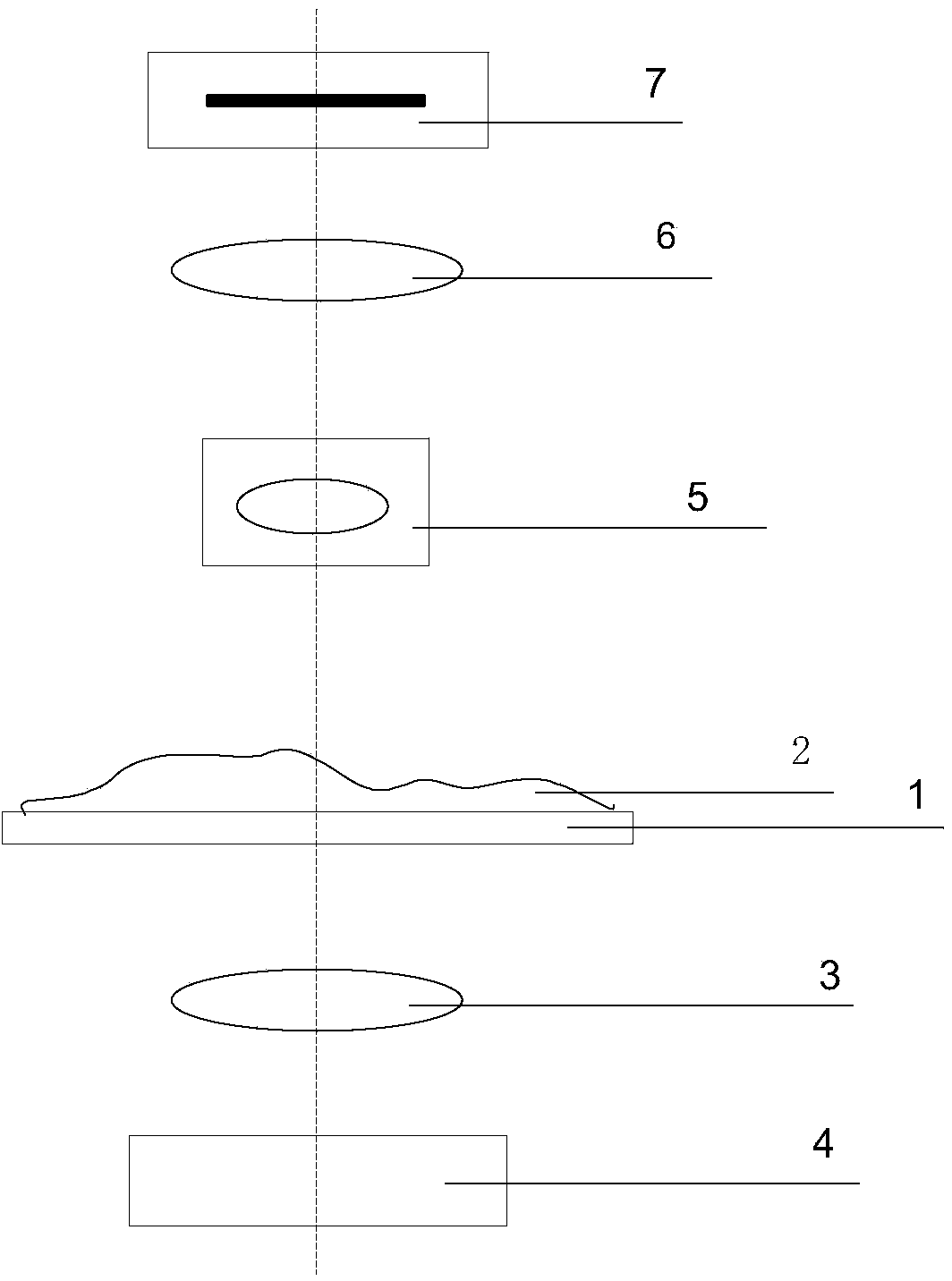
[0044] Step 1: Prepare as attached figure 1 The image acquisition device of the shown structure, wherein, 1 represents a platform, 2 represents a pathological sliced ​​tissue, 3 represents a condenser, 4 represents a light source, 5 represents an objective lens, 6 represents a filter, and 7 represents a camera;

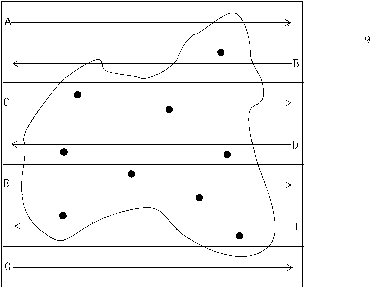
[0045] Step 2: Select representative points on the pathological slice tissue, focus on their focal points, calculate the focal length of each representative point, and fit a rough virtual plane by the least square method, among which, as attached figure 2 The 9 shown indicates the focus;

[0046] Step 3: As attached figure 2 As shown, the first scanning line A of the XYZ axis is set as its trajectory, in which the pathological slice tissue is divided into seven lines A, B, C, D, E, F, and G for scanning, and th...

Embodiment 2

[0053](1) A method for fast and accurate focus scanning of pathological section tissue based on an image acquisition device, the steps of which include:

[0054] Step 1: Prepare as attached Figure 5 The image acquisition device of the shown structure;

[0055] Step 2: Select representative points on the pathological slice tissue, focus on their focal points, calculate the focal length of each representative point, and fit a rough virtual plane;

[0056] Step 3: Set the first scanning line of the XYZ axis as its motion trajectory, and divide the pathological slice tissue into several lines for scanning;

[0057] Step 4: The camera starts to capture the picture. During this process, according to the sharpness trend of the front and rear lines captured by the camera, the movement track of the Z axis is continuously corrected, that is, the focal length is changed;

[0058] Step 5: Complete the scan.

[0059] (2) Principle and operation analysis:

[0060] This embodiment is an...


PUM


Abstract

The invention provides a method for rapidly and accurately focusing and scanning pathological section tissue on the basis of an image collection device. The method comprises steps as follows: (1), the image collection device is prepared; (2), a representative point on the pathological section tissue is selected, a focus point of the representative point is focused, a focal length of each representative point is calculated, and a virtual plane is fit; (3), a first scanning line of X, Y and Z axes is taken as a movement track, and the pathological section tissue is divided into at least two lines for scanning; (4), a camera starts capturing images, the movement track of Z axis is continuously corrected according to the clarity trend of a front line and a rear line obtained by the camera in the process, that is, the focal length is changed; and (5), scanning is completed. The invention further provides a scanner based on the method. By means of the method, a large quantity of focus points is reduced, a rough virtual plane is obtained, and the movement track of the Z axis is continuously corrected according to image data in the scanning process, so that the problems of low focusing speed, inaccuracy and the like are well solved.

Description

technical field [0001] The invention relates to a scanning method, in particular to a method for fast and accurate focusing scanning of pathological slice tissue based on an image acquisition device. The technical field and application products include a digital pathological slice scanner. Background technique [0002] With the continuous development of science and technology, traditional microscopes are constantly being replaced by digital pathological slide scanners. Digital pathological slide scanners not only have the functions of traditional microscopes, but also can digitize and network the pathological resources on slices to realize permanent visualization of data. Storage and synchronous browsing processing not limited by time and space, so that it is widely used in various fields of pathology. [0003] Image acquisition devices under high magnification are generally used in digital pathology slide scanners. The image acquisition devices mainly include light source, ...

Claims


Application Information

Patent Timeline
Patent Type & Authority Applications(China)
IPC IPC(8): G01N21/84
Inventor 刘炳宪谢菊元王焱辉王克惠陈丽桥
Owner KONFOONG BIOTECH INT
Why Eureka
Try Eureka