Organ positioning method based on sectional image reconstruction

A technology of tomographic imaging and positioning method, which is applied in the field of organ positioning, can solve the problems that a person cannot cross the same river twice, and a person maintains a uniform posture.

Inactive Publication Date: 2015-06-03
FUJIAN NORMAL UNIV
View PDF1 Cites 12 Cited by
  • Summary
  • Abstract
  • Description
  • Claims
  • Application Information

AI Technical Summary

Problems solved by technology

In fact this cannot be done, because it is a philosophical question: one cannot cross the same river twice, nor can one maintain the same posture in both tests

Method used


Image

  • Organ positioning method based on sectional image reconstruction
  • Organ positioning method based on sectional image reconstruction
  • Organ positioning method based on sectional image reconstruction

Examples


Embodiment 1

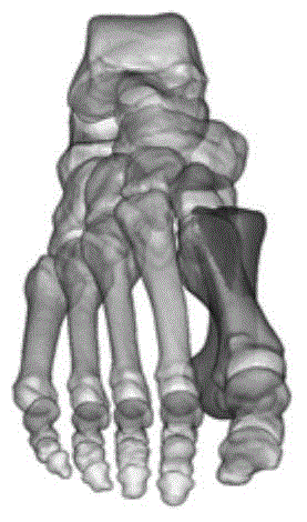
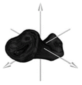
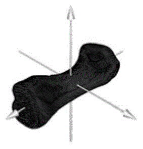
[0039] For human organs, the test position and posture are changed, but this change will not change the shape and structure of the organ. The centroid of human organs is not affected by the scanning position and pose. The main axis of inertia of an object with asymmetric shape and non-uniform mass distribution is also unique. Human organs are asymmetric bodies with non-uniform values. Therefore, the main axis of inertia of human organs is unique. This uniqueness has nothing to do with the test position and posture. According to the invariance of coordinate transformation of geometric bodies, the body coordinate system established based on the main axis of organ inertia in the present invention can realize the positioning of organs.

[0040] Combine below Figure 4 The method adopted in this embodiment is specifically described as follows.

[0041] 1. Scanning equipment

[0042] A tomographic image containing the organ to be located is obtained by scanning such as CT or MRI. ...


PUM


Abstract

The invention discloses an organ positioning method based on sectional image reconstruction. The organ positioning method comprises the following steps: partitioning an organ subjected to sectional image reconstruction into a set of body infinitesimals, obtaining the mass center of the organ according to the coordinate and the density of each infinitesimal in an image, and establishing a body coordinate system that the origin of the coordinate is located on the mass center of the organ, thereby positioning the organ; calculating an inertia principal axis of the organ that all inertia products are zero simultaneously, and positioning the posture of the organ by taking the inertia principal axis of the organ as the coordinate axis of the organ body coordinate system. By virtue of the characteristic that the mass center and the inertia principal axis of the organ are unique, not only is the position of the organ based on sectional image reconstruction quantitatively described, but also the posture of the organ can be quantitatively described, quantification of organ positioning of sectional image reconstruction can be achieved, and the theoretical system of sectional image quantitative analysis can be completed.

Description

technical field [0001] The invention relates to the field of medical tomographic image processing, in particular to an organ positioning method based on tomographic image reconstruction. Background technique [0002] Choosing a reference system and establishing a coordinate system are the prerequisites for describing organ morphology and predicting organ changes. Establishing an organ body coordinate system that has nothing to do with the test position and posture is the premise of effectively helping doctors achieve clinical diagnosis, surgical planning, radiotherapy planning, and pathological change tracking. [0003] Usually, the scanning axis of the instrument is fixed, that is, the coordinate system of the scanning field is fixed. In this way, the body coordinate system of the organ is determined by the position and posture of the subject during the test. Regrettably, there is no way to make the subject maintain the same position and posture in different tests, and the...

Claims


Application Information

Patent Timeline
Patent Type & Authority Applications(China)
IPC IPC(8): G06T7/00G06T7/60
Inventor 范毅方
Owner FUJIAN NORMAL UNIV
Why Eureka
Try Eureka