Chest surgery imaging examination operating table

A technology for thoracic surgery and operating tables, applied in the field of thoracic surgery imaging examination operating tables, can solve problems such as increasing the difficulty of medical staff work, threatening the safety of patients, secondary injuries of patients, etc., to achieve complete functions, reduce work difficulty, and easy to use Effect

Inactive Publication Date: 2015-06-24
刘宝清 +3
View PDF5 Cites 8 Cited by
  • Summary
  • Abstract
  • Description
  • Claims
  • Application Information

AI Technical Summary

Problems solved by technology

[0002] When medical personnel perform thoracic surgery examinations on patients, due to the damaged chest of thoracic surgery patients and the fragility of the injured parts, they cannot perform large movements. The traditional method is to first perform radiological diagnosis on patients and then transfer them to the operating ward for surgical treatment. If an accident occurs during the transfer process, it will cause secondary injury to the patient, which can easily threaten the life safety of the patient and greatly increase the difficulty of the work of the medical staff.

Method used


Image

  • Chest surgery imaging examination operating table
  • Chest surgery imaging examination operating table
  • Chest surgery imaging examination operating table

Examples


Embodiment Construction

[0026] The imaging examination operating table for thoracic surgery of the present invention will be described in detail below with reference to the accompanying drawings.

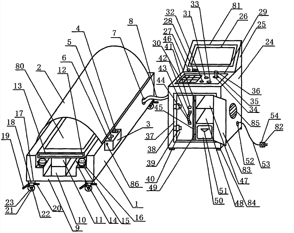
[0027] Such as figure 1 As shown, the thoracic surgery image examination operating table of the present invention includes an examination table main body 1, a radiological inspection cover 2 is arranged on the examination table main body 1, an examination table controller 3 is arranged on the right side of the examination table main body 1, and the examination table The controller 3 is provided with a power switch 4, the front side of the power switch 4 is provided with a mode button 5, the front side of the mode button 5 is provided with a first control button 6, and the rear side of the examination table controller 3 is provided with a control line nozzle 7, and the control A control line pipe 8 is arranged in the line pipe port 7 .

[0028] Such as figure 1 As shown, a U-shaped groove 9 is arranged in...


PUM


Abstract

The invention relates to a chest surgery imaging examination operating table, and belongs to the technical field of medical apparatuses and instruments. The chest surgery imaging examination operating table comprises an examination table body, a radioscopy examination cover is arranged on the examination table body, an examination table controller is arranged on the right lateral face of the examination table body, a power switch is arranged on the examination table controller, a mode button is arranged on the front side of the power switch, a first control button is arranged on the front side of the mode button, a control line pipe opening is formed in the back side of the examination table controller, and a control line pipe is arranged in the control line pipe opening. The chest surgery imaging examination operating table is complete in function and convenient to use; when chest surgery examination is carried out, time and labor are saved, the chest surgery imaging examination operating table is scientific, convenient, rapid, safe, efficient, intelligent and accurate, and the work difficulty of medical workers is reduced.

Description

technical field [0001] The invention belongs to the technical field of medical devices, and in particular relates to an image examination operating table for thoracic surgery. Background technique [0002] When medical personnel perform thoracic surgery examinations on patients, due to the damaged chest of thoracic surgery patients and the fragility of the injured parts, they cannot perform large movements. The traditional method is to first perform radiological diagnosis on patients and then transfer them to the operating ward for surgical treatment. If an accident occurs during the transfer process, it will cause secondary injury to the patient, which can easily threaten the life safety of the patient and greatly increase the work difficulty of the medical staff. Contents of the invention [0003] The purpose of the present invention is to provide a thoracic surgery imaging examination operating table which is time-saving, scientific and convenient, safe and efficient, i...

Claims


Application Information

Patent Timeline
Patent Type & Authority Applications(China)
IPC IPC(8): A61G13/00A61G13/10A61B6/00
Inventor 戴伟孟庆萍刘思爱曹丽萍孔斌刘思梅张莉
Owner 刘宝清
Why Eureka
Try Eureka