Application system for subdural hematoma volume calculation

A volume calculation and application system technology, applied in the field of medical application, can solve the problems of high cost, inaccuracy, and time-consuming, and achieve the effects of accurate measurement, convenient operation, and high calculation accuracy

Active Publication Date: 2015-07-15
SHENYANG AEROSPACE UNIVERSITY
5 Cites 0 Cited by

AI-Extracted Technical Summary

Problems solved by technology

The Tada formula method is more accurate for the measurement of hematoma with small bleeding volume and ellipsoidal shape, but it is not accurate for the measurement of large bleeding volume, irregular shape, intraventricular hemorrhage, and subdural hematoma; althoug...

Abstract

In order to overcome the defects in the prior art, the invention aims at providing an application system for subdural hematoma volume calculation with the advantages that the intracranial hematoma volume can be accurately measured, the time consumption is low, and the money consumption is low. The application system has the technical scheme that the application system for subdural hematoma volume calculation comprises an information measurement input module, a hematoma volume calculation module and a hematoma volume output module. A control method is characterized in that a VB (visual basic) program is compiled on the basis of a polygon formula, points are drawn on a CT (computed tomography) image, each layer of CT approximates to a polygon, then, the area of each layer of CT image is calculated by utilizing the compiled program, and the volume of each layer is further calculated through multiplying the area by the thickness and the CT image proportion,.

Application Domain

Image analysisComputerised tomographs +1

Technology Topic

Computed tomographyVisual Basic +4

Image

  • Application system for subdural hematoma volume calculation
  • Application system for subdural hematoma volume calculation
  • Application system for subdural hematoma volume calculation

Examples

  • Experimental program(1)

Example Embodiment

[0020] Example 1:
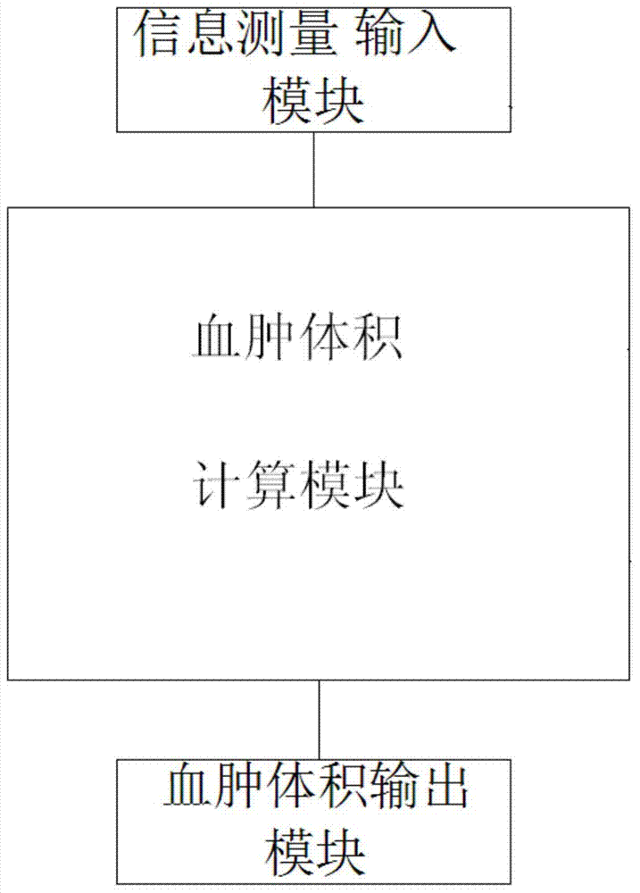
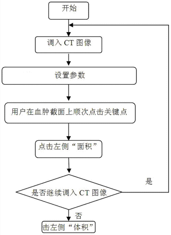
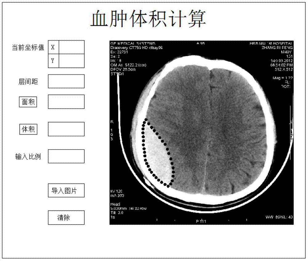
[0021] Such as Figure 1 to Figure 3 Shown: An application system for subdural hematoma volume calculation, including an information measurement input module, a hematoma volume calculation module, and a hematoma volume output module. The information measurement input module is used to complete the measurement of related parameters and integrate it Input to the hematoma volume calculation module, the hematoma volume calculation module is used to complete the hematoma volume calculation, and the hematoma volume output module outputs the hematoma volume. The described hematoma volume calculation module can be composed of a computer and VB software. Its control method is based on the polygon formula method to write a VB program. By drawing points on the CT image, each layer of CT is approximated as a polygon, and then the written program is used to calculate each The area of ​​the slice CT image is calculated by multiplying the area by the thickness and the CT image ratio to calculate the volume of each slice. The formula used in this method:
[0022] Calculation formula of CT image area of ​​each layer:
[0023] Hematoma volume calculation formula:
[0024] Where c is the number of layers and h is the layer spacing.
[0025] Use VB to write the program. When the operator clicks on the key points on each CT scan image, the program will record the coordinates of a series of key points in sequence, and automatically connect the last key point and the first key point with a straight line. Reflected in the program as (x k+1 , Y k+1 )=(x 1 , Y 1 ), these coordinates provide raw data for measuring the volume, and the specific implementation is as follows:
[0026] 1. Establish a coordinate system in the VB interface: establish a program through the coordinate system to define the coordinate accuracy and size of the coordinate system;
[0027] 2. In the VB interface, double-click the add button and display box command, add the following buttons and displays on the interface in turn, and then set the control program of each button:
[0028] (1) The "Import Picture" button corresponds to the "Program for Writing Related Picture Information". When you click this button, you can select the picture that needs to be imported;
[0029] (2) "Current coordinate value" corresponds to "Display the coordinates of the point clicked by the mouse". When you click a point in the interface with the left mouse button, the "Current coordinate value" window will display the values ​​of x and y, and Assign the values ​​of x and y to the formula S j = 1 2 X i = 1 n ( x i y i + 1 - x i + 1 y i ) X in i , Y i;
[0030] (3) "Area" button corresponds to "Area solving formula ", when you stop clicking on the point in the CT image on the interface, click the "Area" button to get the area of ​​the shadow on the CT image, and assign this value to the volume solving formula S in j;
[0031] (4) The "Layer Spacing" window corresponds to the "Parameter Assignment Procedure". When entering the value, you can give the formula V = k · h · X j = 1 c S j H assignment in;
[0032] (5) The "input ratio" window corresponds to the "parameter assignment procedure", when a certain value is input, this value is assigned to the volume formula V = k · h · X j = 1 c S j K in
[0033] (6) The "Volume" button corresponds to the "Volume Solving Formula", click the "Volume" button to get the hematoma volume value;
[0034] (7) The "Clear" button corresponds to the "Clear instruction in the program". Click this button to clear the information in the above window.
[0035] The operating steps of the hematoma volume calculation system are as follows:
[0036] (1) Import CT images;
[0037] (2) To set the parameters, enter the ratio k and the layer spacing h. The ratio and layer spacing are all known and determined values. The parameters can be determined according to the settings of the CT machine itself;
[0038] (3) Click the key points in sequence on the hematoma section;
[0039] (4) Click the "Area" button in the VB interface to output the area;
[0040] (5) Follow steps (1)-(4) to process all CT images in sequence
[0041] (6) Click the "Volume" button in the VB interface to get the hematoma volume.

PUM

no PUM

Description & Claims & Application Information

We can also present the details of the Description, Claims and Application information to help users get a comprehensive understanding of the technical details of the patent, such as background art, summary of invention, brief description of drawings, description of embodiments, and other original content. On the other hand, users can also determine the specific scope of protection of the technology through the list of claims; as well as understand the changes in the life cycle of the technology with the presentation of the patent timeline. Login to view more.

Similar technology patents

3D Gauss-FFT based gravity field forward modeling method

InactiveCN108984939AImprove calculation accuracyImprove the accuracy of forward modeling
Owner:CENT SOUTH UNIV

Classification and recommendation of technical efficacy words

  • easy to operate
  • Improve calculation accuracy

Lancet device

InactiveUS7303573B2inexpensively mass-producedeasy to operate
Owner:ABBOTT DIABETES CARE INC

Rapid and non-invasive optical detection of internal bleeding

InactiveUS20050065436A1easy to operaterapid and accurate result
Owner:HO WINSTON ZONH +2

Aircraft conflict relief method and device

Owner:北京福瑞航行科技有限公司
Why Eureka
Try Eureka